ARG57063
anti-VTA1 antibody [14G10]
anti-VTA1 antibody [14G10] for Flow cytometry,ICC/IF,Western blot and Human
Overview
| Product Description | Mouse Monoclonal antibody [14G10] recognizes VTA1 |
|---|---|
| Tested Reactivity | Hu |
| Tested Application | FACS, ICC/IF, WB |
| Host | Mouse |
| Clonality | Monoclonal |
| Clone | 14G10 |
| Isotype | IgG1, kappa |
| Target Name | VTA1 |
| Antigen Species | Human |
| Immunogen | Recombinant fragment around aa. 1-307 of Human VTA1. |
| Conjugation | Un-conjugated |
| Alternate Names | DRG1; HSPC228; Vacuolar protein sorting-associated protein VTA1 homolog; SKD1-binding protein 1; LYST-interacting protein 5; C6orf55; LIP5; SBP1; My012; Dopamine-responsive gene 1 protein; DRG-1 |
Application Instructions
| Application Suggestion |
|
||||||||
|---|---|---|---|---|---|---|---|---|---|
| Application Note | * The dilutions indicate recommended starting dilutions and the optimal dilutions or concentrations should be determined by the scientist. |
Properties
| Form | Liquid |
|---|---|
| Purification | Purification with Protein A. |
| Buffer | PBS (pH 7.4), 0.02% Sodium azide and 10% Glycerol. |
| Preservative | 0.02% Sodium azide |
| Stabilizer | 10% Glycerol |
| Concentration | 1 mg/ml |
| Storage Instruction | For continuous use, store undiluted antibody at 2-8°C for up to a week. For long-term storage, aliquot and store at -20°C. Storage in frost free freezers is not recommended. Avoid repeated freeze/thaw cycles. Suggest spin the vial prior to opening. The antibody solution should be gently mixed before use. |
| Note | For laboratory research only, not for drug, diagnostic or other use. |
Bioinformation
| Database Links |
Swiss-port # Q9NP79 Human Vacuolar protein sorting-associated protein VTA1 homolog |
|---|---|
| Gene Symbol | VTA1 |
| Gene Full Name | vesicle (multivesicular body) trafficking 1 |
| Background | C6ORF55 encodes a protein involved in trafficking of the multivesicular body, an endosomal compartment involved in sorting membrane proteins for degradation in lysosomes (Ward et al., 2005 [PubMed 15644320]).[supplied by OMIM, Mar 2008] |
| Function | Involved in the endosomal multivesicular bodies (MVB) pathway. MVBs contain intraluminal vesicles (ILVs) that are generated by invagination and scission from the limiting membrane of the endosome and mostly are delivered to lysosomes enabling degradation of membrane proteins, such as stimulated growth factor receptors, lysosomal enzymes and lipids. Thought to be a cofactor of VPS4A/B, which catalyzes disassembles membrane-associated ESCRT-III assemblies. Involved in the sorting and down-regulation of EGFR (By similarity). Involved in HIV-1 budding. [UniProt] |
| Calculated MW | 34 kDa |
Images (3) Click the Picture to Zoom In
-
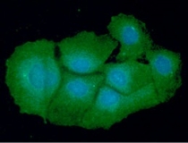
ARG57063 anti-VTA1 antibody [14G10] ICC/IF image
Immunoflorescense: Hep3B cell line stained with ARG57063 anti-VTA1 antibody [14G10] at 1:100 (Green).
DAPI (Blue) for nucleus staining.
-
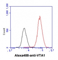
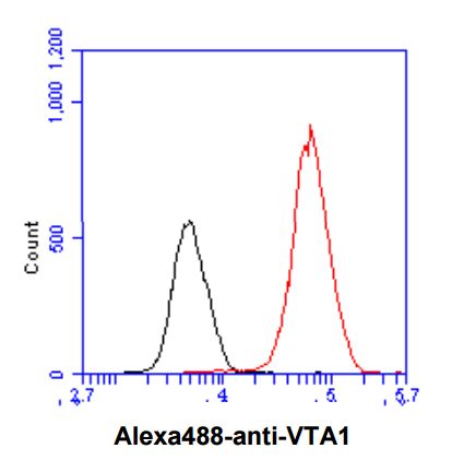
ARG57063 anti-VTA1 antibody [14G10] FACS image
Flow Cytometry: Hep3B cell line stained with ARG57063 anti-VTA1 antibody [14G10] at 2-5 µg for 1x10^6 cells (red line). Secondary antibody: Goat anti-Mouse IgG Alexa fluor 488 conjugate. Isotype control antibody was Mouse IgG (black line).
-
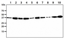
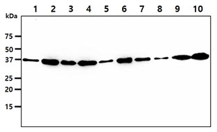
ARG57063 anti-VTA1 antibody [14G10] WB image
Western blot: 40 µg of 1) HepG2 cell lysate, 2) 293T cell lysate, 3) HeLa cell lysate, 4) MCF7 cell lysate, 5) A549 cell lysate, 6) Jurkat cell lysate, 7) K562 cell lysate, 8) LnCaP cell lysate, 9) U937 cell lysate, 10) A431 cell lysate stained with ARG57063 anti-VTA1 antibody [14G10] at 1:1000.