ARG63867
anti-VPS37C antibody
anti-VPS37C antibody for IHC-Formalin-fixed paraffin-embedded sections,Western blot and Human
Signaling Transduction antibody
Overview
| Product Description | Goat Polyclonal antibody recognizes VPS37C |
|---|---|
| Tested Reactivity | Hu |
| Tested Application | IHC-P, WB |
| Host | Goat |
| Clonality | Polyclonal |
| Isotype | IgG |
| Target Name | VPS37C |
| Antigen Species | Human |
| Immunogen | C-ETLKDKTLQELEELQ |
| Conjugation | Un-conjugated |
| Alternate Names | hVps37C; Vacuolar protein sorting-associated protein 37C; ESCRT-I complex subunit VPS37C |
Application Instructions
| Application Suggestion |
|
||||||
|---|---|---|---|---|---|---|---|
| Application Note | WB: Recommend incubate at RT for 1h. IHC-P: Antigen Retrieval: Steam tissue section in Citrate buffer (pH 6.0). * The dilutions indicate recommended starting dilutions and the optimal dilutions or concentrations should be determined by the scientist. |
Properties
| Form | Liquid |
|---|---|
| Purification | Purified from goat serum by ammonium sulphate precipitation followed by antigen affinity chromatography using the immunizing peptide. |
| Buffer | Tris saline (pH 7.3), 0.02% Sodium azide and 0.5% BSA |
| Preservative | 0.02% Sodium azide |
| Stabilizer | 0.5% BSA |
| Concentration | 0.5 mg/ml |
| Storage Instruction | For continuous use, store undiluted antibody at 2-8°C for up to a week. For long-term storage, aliquot and store at -20°C or below. Storage in frost free freezers is not recommended. Avoid repeated freeze/thaw cycles. Suggest spin the vial prior to opening. The antibody solution should be gently mixed before use. |
| Note | For laboratory research only, not for drug, diagnostic or other use. |
Bioinformation
| Database Links |
Swiss-port # A5D8V6 Human Vacuolar protein sorting-associated protein 37C |
|---|---|
| Background | VPS37C is a subunit of ESCRT-I (endosomal sorting complex required for transport I), a complex in the class E vacuolar protein sorting (VPS) pathway required for sorting ubiquitinated transmembrane proteins into internal vesicles of multivesicular bodies (Eastman et al., 2005 [PubMed 15509564]).[supplied by OMIM, Mar 2008] |
| Research Area | Signaling Transduction antibody |
| Calculated MW | 39 kDa |
| PTM | Phosphorylated by TBK1. |
Images (3) Click the Picture to Zoom In
-
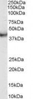
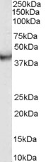
ARG63867 anti-VPS37C antibody WB image
Western Blot: human duodenum lysate (35 µg protein in RIPA buffer) stained with ARG63867 anti-VPS37C antibody at 0.03g/ml dilution.
-
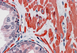
ARG63867 anti-VPS37C antibody IHC-P image
Immunohistochemistry: Paraffin-embedded Human breast tissue. Antigen Retrieval: Steam tissue section in Citrate buffer (pH 6.0). The tissue section was stained with ARG63867 anti-VPS37C antibody at 3.75 µg/ml dilution followed by AP-staining.
-
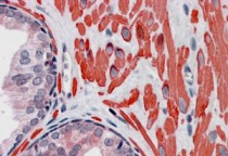
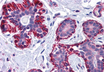
ARG63867 anti-VPS37C antibody IHC-P image
Immunohistochemistry: paraffin embedded Human Prostate. (Steamed antigen retrieval with citrate buffer pH 6) stained with ARG63867 anti-VPS37C antibody at 3.8 µg/ml dilution followed by AP-staining.