ARG52467
anti-VGAT antibody
anti-VGAT antibody for Western blot and Rat
Neuroscience antibody
Overview
| Product Description | Rabbit Polyclonal antibody recognizes VGAT |
|---|---|
| Tested Reactivity | Rat |
| Predict Reactivity | Bov, Dog, NHuPrm |
| Tested Application | WB |
| Host | Rabbit |
| Clonality | Polyclonal |
| Isotype | IgG |
| Target Name | VGAT |
| Antigen Species | Rat |
| Immunogen | Synthetic peptide corresponding to amino acid residues from the N-terminal region conjugated to KLH |
| Conjugation | Un-conjugated |
| Alternate Names | Vesicular GABA transporter; GABA and glycine transporter; Vesicular inhibitory amino acid transporter; VIAAT; hVIAAT; VGAT; Solute carrier family 32 member 1 |
Application Instructions
| Application Suggestion |
|
||||
|---|---|---|---|---|---|
| Application Note | Specific for the ~53k VGAT protein. * The dilutions indicate recommended starting dilutions and the optimal dilutions or concentrations should be determined by the scientist. |
Properties
| Form | Liquid |
|---|---|
| Purification | Affinity Purified |
| Buffer | 10 mM HEPES (pH 7.5), 150 mM NaCl, 0.1 mg/ml BSA and 50% Glycerol |
| Stabilizer | 0.1 mg/ml BSA, 50% Glycerol |
| Storage Instruction | For continuous use, store undiluted antibody at 2-8°C for up to a week. For long-term storage, aliquot and store at -20°C. Storage in frost free freezers is not recommended. Avoid repeated freeze/thaw cycles. Suggest spin the vial prior to opening. The antibody solution should be gently mixed before use. |
| Note | For laboratory research only, not for drug, diagnostic or other use. |
Bioinformation
| Database Links |
Swiss-port # O35458 Rat Vesicular inhibitory amino acid transporter |
|---|---|
| Gene Symbol | SLC32A1 |
| Gene Full Name | solute carrier family 32 (GABA vesicular transporter), member 1 |
| Background | The Vesicular GABA Amino Acid Transporter (VGAT) is responsible for transport of the inhibitory neurotransmitter into synaptic vesicles(McIntire et al., 1997). The VGAT protein (also known as the Vesicular Inhibitory Amino Aid Transporter or VIAAT) is expressed in synaptic vesicles of both glycine and GABAergic synapses throughout the CNS (Chaudhry et al., 1998). Expression of the VGAT protein changes during development and also in response to patterns of neuronal activity (De et al., 2005). |
| Highlight | Related products: Anti-Rabbit IgG secondary antibodies; Related news: Neuronal Development Marker |
| Research Area | Neuroscience antibody |
| Calculated MW | 57 kDa |
Images (1) Click the Picture to Zoom In
-
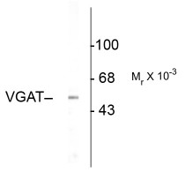
ARG52467 anti-VGAT antibody WB image
Western Blot: rat olfactory bulb lysate showing specific immunolabeling of the ~53k VGAT protein stained with VGAT antibody (ARG52467).


