ARG51821
anti-VEGFR2 phospho (Tyr1059) antibody
anti-VEGFR2 phospho (Tyr1059) antibody for Western blot and Human,Mouse
Cancer antibody; Cell Biology and Cellular Response antibody; Controls and Markers antibody; Developmental Biology antibody; Metabolism antibody; Microbiology and Infectious Disease antibody; Signaling Transduction antibody
Overview
| Product Description | Rabbit Polyclonal antibody recognizes VEGFR2 phospho (Tyr1059) |
|---|---|
| Tested Reactivity | Hu, Ms |
| Tested Application | WB |
| Host | Rabbit |
| Clonality | Polyclonal |
| Isotype | IgG |
| Target Name | VEGFR2 |
| Antigen Species | Human |
| Immunogen | Peptide sequence around phosphorylation site of tyrosine 1059 (P-D-Y(p)-V-R)derived from Human VEGFR2. |
| Conjugation | Un-conjugated |
| Alternate Names | FLK1; VEGFR; CD antigen CD309; FLK-1; Fetal liver kinase 1; VEGFR2; Vascular endothelial growth factor receptor 2; VEGFR-2; CD309; Kinase insert domain receptor; EC 2.7.10.1; Protein-tyrosine kinase receptor flk-1; KDR |
Application Instructions
| Application Suggestion |
|
||||
|---|---|---|---|---|---|
| Application Note | * The dilutions indicate recommended starting dilutions and the optimal dilutions or concentrations should be determined by the scientist. |
Properties
| Form | Liquid |
|---|---|
| Purification | Antibodies were produced by immunizing rabbits with KLH-conjugated synthetic phosphopeptide. Antibodies were purified by affinity-chromatography using epitope-specific phosphopeptide. In addition, non-phospho specific antibodies were removed by chromatogramphy using non-phosphopeptide. |
| Buffer | PBS (without Mg2+ and Ca2+, pH 7.4), 150mM NaCl, 0.02% Sodium azide and 50% Glycerol. |
| Preservative | 0.02% Sodium azide |
| Stabilizer | 50% Glycerol |
| Concentration | 1 mg/ml |
| Storage Instruction | For continuous use, store undiluted antibody at 2-8°C for up to a week. For long-term storage, aliquot and store at -20°C. Storage in frost free freezers is not recommended. Avoid repeated freeze/thaw cycles. Suggest spin the vial prior to opening. The antibody solution should be gently mixed before use. |
| Note | For laboratory research only, not for drug, diagnostic or other use. |
Bioinformation
| Database Links |
Swiss-port # P35918 Mouse Vascular endothelial growth factor receptor 2 Swiss-port # P35968 Human Vascular endothelial growth factor receptor 2 |
|---|---|
| Gene Symbol | KDR |
| Gene Full Name | kinase insert domain receptor |
| Background | Receptor for VEGF or VEGFC. Has a tyrosine-protein kinase activity. The VEGF-kinase ligand/receptor signaling system plays a key role in vascular development and regulation of vascular permeability. In case of HIV-1 infection, the interaction with extracellular viral Tat protein seems to enhance angiogenesis in Kaposi's sarcoma lesions |
| Function | Tyrosine-protein kinase that acts as a cell-surface receptor for VEGFA, VEGFC and VEGFD. Plays an essential role in the regulation of angiogenesis, vascular development, vascular permeability, and embryonic hematopoiesis. Promotes proliferation, survival, migration and differentiation of endothelial cells. Promotes reorganization of the actin cytoskeleton. Isoforms lacking a transmembrane domain, such as isoform 2 and isoform 3, may function as decoy receptors for VEGFA, VEGFC and/or VEGFD. Isoform 2 plays an important role as negative regulator of VEGFA- and VEGFC-mediated lymphangiogenesis by limiting the amount of free VEGFA and/or VEGFC and preventing their binding to FLT4. Modulates FLT1 and FLT4 signaling by forming heterodimers. Binding of vascular growth factors to isoform 1 leads to the activation of several signaling cascades. Activation of PLCG1 leads to the production of the cellular signaling molecules diacylglycerol and inositol 1,4,5-trisphosphate and the activation of protein kinase C. Mediates activation of MAPK1/ERK2, MAPK3/ERK1 and the MAP kinase signaling pathway, as well as of the AKT1 signaling pathway. Mediates phosphorylation of PIK3R1, the regulatory subunit of phosphatidylinositol 3-kinase, reorganization of the actin cytoskeleton and activation of PTK2/FAK1. Required for VEGFA-mediated induction of NOS2 and NOS3, leading to the production of the signaling molecule nitric oxide (NO) by endothelial cells. Phosphorylates PLCG1. Promotes phosphorylation of FYN, NCK1, NOS3, PIK3R1, PTK2/FAK1 and SRC. [UniProt] |
| Research Area | Cancer antibody; Cell Biology and Cellular Response antibody; Controls and Markers antibody; Developmental Biology antibody; Metabolism antibody; Microbiology and Infectious Disease antibody; Signaling Transduction antibody |
| Calculated MW | 152 kDa |
| PTM | N-glycosylated. Ubiquitinated. Tyrosine phosphorylation of the receptor promotes its poly-ubiquitination, leading to its degradation via the proteasome or lysosomal proteases. Autophosphorylated on tyrosine residues upon ligand binding. Autophosphorylation occurs in trans, i.e. one subunit of the dimeric receptor phosphorylates tyrosine residues on the other subunit. Phosphorylation at Tyr-951 is important for interaction with SH2D2A/TSAD and VEGFA-mediated reorganization of the actin cytoskeleton. Phosphorylation at Tyr-1175 is important for interaction with PLCG1 and SHB. Phosphorylation at Tyr-1214 is important for interaction with NCK1 and FYN. Dephosphorylated by PTPRB. Dephosphorylated by PTPRJ at Tyr-951, Tyr-996, Tyr-1054, Tyr-1059, Tyr-1175 and Tyr-1214. The inhibitory disulfide bond between Cys-1024 and Cys-1045 may serve as a specific molecular switch for H(2)S-induced modification that regulates VEGFR2 function. |
Images (1) Click the Picture to Zoom In
-
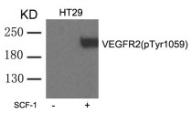
ARG51821 anti-VEGFR2 phospho (Tyr1059) antibody WB image
Western blot: Extracts from HT29 cells untreated or treated with SCF-1 stained with ARG51821 anti-VEGFR2 phospho (Tyr1059) antibody.


