ARG20567
anti-VE Cadherin phospho (Tyr685) antibody
anti-VE Cadherin phospho (Tyr685) antibody for Western blot and Human
Cancer antibody; Controls and Markers antibody; Signaling Transduction antibody
Overview
| Product Description | Rabbit Polyclonal antibody recognizes VE Cadherin phospho (Tyr685) |
|---|---|
| Tested Reactivity | Hu |
| Predict Reactivity | Ms, Rat |
| Tested Application | WB |
| Specificity | The purified antibody detects a ~140 kDa band corresponding to VE-cadherin in western blots of human endothelial cells treated with pervanadate, and this band is not detected in untreated cells or after alkaline phosphatase treatment. This sequence is not conserved in other cadherins. |
| Host | Rabbit |
| Clonality | Polyclonal |
| Isotype | IgG |
| Target Name | VE Cadherin |
| Antigen Species | Human |
| Immunogen | Phosphospecific peptide (coupled to carrier protein) around Tyr685 of Human VE-cadherin. |
| Conjugation | Un-conjugated |
| Alternate Names | 7B4 antigen; 7B4; Cadherin-5; VE-cadherin; CD144; CD antigen CD144; Vascular endothelial cadherin |
Application Instructions
| Application Suggestion |
|
||||
|---|---|---|---|---|---|
| Application Note | WB: Antibody is suggested to be diluted in 5% skimmed milk/Tris buffer with 0.04% Tween20 and incubated for 1 hour at room temperature. * The dilutions indicate recommended starting dilutions and the optimal dilutions or concentrations should be determined by the scientist. |
||||
| Observed Size | 140 kDa |
Properties
| Purification | This antibody was pre-adsorbed with an unrelated phospho-tyrosine peptide and unphosphorylated VE-cadherin (Tyr685) peptide then affinity purified with immunogen peptide. |
|---|---|
| Buffer | PBS, 50% Glycerol, 1 mg/ml BSA and 0.05% Sodium azide |
| Preservative | 0.05% Sodium azide |
| Stabilizer | 50% Glycerol, 1 mg/ml BSA |
| Storage Instruction | For continuous use, store undiluted antibody at 2-8°C for up to a week. For long-term storage, aliquot and store at -20°C. Storage in frost free freezers is not recommended. Avoid repeated freeze/thaw cycles. Suggest spin the vial prior to opening. The antibody solution should be gently mixed before use. |
| Note | For laboratory research only, not for drug, diagnostic or other use. |
Bioinformation
| Database Links | |
|---|---|
| Gene Symbol | CDH5 |
| Gene Full Name | cadherin 5, type 2 (vascular endothelium) |
| Background | This gene is a classical cadherin from the cadherin superfamily and is located in a six-cadherin cluster in a region on the long arm of chromosome 16 that is involved in loss of heterozygosity events in breast and prostate cancer. The encoded protein is a calcium-dependent cell-cell adhesion glycoprotein comprised of five extracellular cadherin repeats, a transmembrane region and a highly conserved cytoplasmic tail. Functioning as a classic cadherin by imparting to cells the ability to adhere in a homophilic manner, the protein may play an important role in endothelial cell biology through control of the cohesion and organization of the intercellular junctions. An alternative splice variant has been described but its full length sequence has not been determined. [provided by RefSeq, Jul 2008] |
| Function | Cadherins are calcium-dependent cell adhesion proteins. They preferentially interact with themselves in a homophilic manner in connecting cells; cadherins may thus contribute to the sorting of heterogeneous cell types. This cadherin may play a important role in endothelial cell biology through control of the cohesion and organization of the intercellular junctions. It associates with alpha-catenin forming a link to the cytoskeleton. Acts in concert with KRIT1 to establish and maintain correct endothelial cell polarity and vascular lumen. These effects are mediated by recruitment and activation of the Par polarity complex and RAP1B. Required for activation of PRKCZ and for the localization of phosphorylated PRKCZ, PARD3, TIAM1 and RAP1B to the cell junction. [UniProt] |
| Research Area | Cancer antibody; Controls and Markers antibody; Signaling Transduction antibody |
| Calculated MW | 88 kDa (unmodified); 90 - 140 kDa (glycosylated) |
| PTM | Phosphorylated on tyrosine residues by KDR/VEGFR-2. Dephosphorylated by PTPRB (By similarity). O-glycosylated. |
Images (1) Click the Picture to Zoom In
-
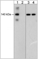
ARG20567 anti-VE Cadherin phospho (Tyr685) antibody WB image
Western blot: Human umbilical vein endothelial cells stimulated with pervanadate (1 mM) for 30 min. then the blots were untreated (Lanes 1 & 3) or treated with alkaline phosphatase (Lanes 2 & 4). The blots were stained with ARG20567 anti-VE Cadherin phospho (Tyr685) antibody (Lanes 1 & 2) or Mouse monoclonal anti-VE-cadherin antibody (Lanes 3 & 4).


