ARG63455
anti-VDAC2 antibody
anti-VDAC2 antibody for IHC-Formalin-fixed paraffin-embedded sections,Western blot and Human,Mouse,Rat,Pig
Metabolism antibody; Signaling Transduction antibody

Overview
| Product Description | Goat Polyclonal antibody recognizes VDAC2 |
|---|---|
| Tested Reactivity | Hu, Ms, Rat, Pig |
| Predict Reactivity | Cow, Dog |
| Tested Application | IHC-P, WB |
| Host | Goat |
| Clonality | Polyclonal |
| Isotype | IgG |
| Target Name | VDAC2 |
| Antigen Species | Human |
| Immunogen | C-GHKVGLALELEA |
| Conjugation | Un-conjugated |
| Alternate Names | Voltage-dependent anion-selective channel protein 2; hVDAC2; POR; VDAC-2; Outer mitochondrial membrane protein porin 2 |
Application Instructions
| Application Suggestion |
|
||||||
|---|---|---|---|---|---|---|---|
| Application Note | WB: Recommend incubate at RT for 1h. IHC-P: Antigen Retrieval: Steam tissue section in Citrate buffer (pH 6.0). * The dilutions indicate recommended starting dilutions and the optimal dilutions or concentrations should be determined by the scientist. |
Properties
| Form | Liquid |
|---|---|
| Purification | Purified from goat serum by ammonium sulphate precipitation followed by antigen affinity chromatography using the immunizing peptide. |
| Buffer | Tris saline (pH 7.3), 0.02% Sodium azide and 0.5% BSA |
| Preservative | 0.02% Sodium azide |
| Stabilizer | 0.5% BSA |
| Concentration | 0.5 mg/ml |
| Storage Instruction | For continuous use, store undiluted antibody at 2-8°C for up to a week. For long-term storage, aliquot and store at -20°C or below. Storage in frost free freezers is not recommended. Avoid repeated freeze/thaw cycles. Suggest spin the vial prior to opening. The antibody solution should be gently mixed before use. |
| Note | For laboratory research only, not for drug, diagnostic or other use. |
Bioinformation
| Database Links | |
|---|---|
| Background | This gene encodes a member of the voltage-dependent anion channel pore-forming family of proteins that are considered the main pathway for metabolite diffusion across the mitochondrial outer membrane. The encoded protein is also thought to be involved in the mitochondrial apoptotic pathway via regulation of BCL2-antagonist/killer 1 protein activity. Pseudogenes have been identified on chromosomes 1, 2, 12 and 21, and alternative splicing results in multiple transcript variants. [provided by RefSeq, May 2010] |
| Research Area | Metabolism antibody; Signaling Transduction antibody |
| Calculated MW | 32 kDa |
| PTM | Ubiquitinated by PRKN during mitophagy, leading to its degradation and enhancement of mitophagy. Deubiquitinated by USP30. |
Images (2) Click the Picture to Zoom In
-
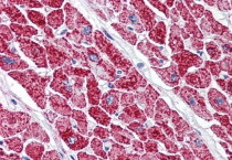
ARG63455 anti-VDAC2 antibody IHC-P image
Immunohistochemistry: Paraffin-embedded Human heart tissue. Antigen Retrieval: Steam tissue section in Citrate buffer (pH 6.0). The tissue section was stained with ARG63455 anti-VDAC2 antibody at 5 µg/ml dilution followed by AP-staining.
-

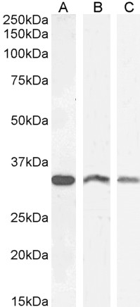
ARG63455 anti-VDAC2 antibody WB image
Western blot: 35 µg of Human (A), Mouse (B) and Rat (C) brain lysates (in RIPA buffer) stained with ARG63455 anti-VDAC2 antibody at 0.3 µg/ml (A) and 1 µg/ml (B, C) dilutions and incubated at RT for 1 hour.

Customer's Feedback
Good
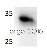
Review for anti-VDAC2 antibody
Application:WB
Sample:HT29
Sample Loading Amount:30 µg
Primary Antibody Dilution Factor:1:500
Primary Antibody Incubation Time:overnight
Primary Antibody Incubation Temperature:4 ºC


