ARG59189
anti-USP7 / HAUSP antibody
anti-USP7 / HAUSP antibody for Flow cytometry,ICC/IF,IHC-Formalin-fixed paraffin-embedded sections,Western blot and Human,Mouse,Rat
Overview
| Product Description | Rabbit Polyclonal antibody recognizes USP7 / HAUSP |
|---|---|
| Tested Reactivity | Hu, Ms, Rat |
| Tested Application | FACS, ICC/IF, IHC-P, WB |
| Host | Rabbit |
| Clonality | Polyclonal |
| Isotype | IgG |
| Target Name | USP7 / HAUSP |
| Antigen Species | Human |
| Immunogen | Recombinant protein corresponding to L258-D483 of Human USP7 / HAUSP. |
| Conjugation | Un-conjugated |
| Alternate Names | Ubiquitin-specific-processing protease 7; Ubiquitin carboxyl-terminal hydrolase 7; HAUSP; Herpesvirus-associated ubiquitin-specific protease; Deubiquitinating enzyme 7; TEF1; Ubiquitin thioesterase 7; EC 3.4.19.12 |
Application Instructions
| Application Suggestion |
|
||||||||||
|---|---|---|---|---|---|---|---|---|---|---|---|
| Application Note | IHC-P: Antigen Retrieval: Heat mediation was performed in Citrate buffer (pH 6.0) for 20 min. * The dilutions indicate recommended starting dilutions and the optimal dilutions or concentrations should be determined by the scientist. |
Properties
| Form | Liquid |
|---|---|
| Purification | Affinity purification with immunogen. |
| Buffer | 0.9% NaCl, 0.2% Na2HPO4, 0.05% Sodium azide and 4% Trehalose. |
| Preservative | 0.05% Sodium azide |
| Stabilizer | 4% Trehalose |
| Concentration | 0.5 mg/ml |
| Storage Instruction | For continuous use, store undiluted antibody at 2-8°C for up to a week. For long-term storage, aliquot and store at -20°C or below. Storage in frost free freezers is not recommended. Avoid repeated freeze/thaw cycles. Suggest spin the vial prior to opening. The antibody solution should be gently mixed before use. |
| Note | For laboratory research only, not for drug, diagnostic or other use. |
Bioinformation
| Database Links | |
|---|---|
| Gene Symbol | USP7 |
| Gene Full Name | ubiquitin specific peptidase 7 (herpes virus-associated) |
| Function | Hydrolase that deubiquitinates target proteins such as FOXO4, p53/TP53, MDM2, ERCC6, DNMT1, UHRF1, PTEN and DAXX. Together with DAXX, prevents MDM2 self-ubiquitination and enhances the E3 ligase activity of MDM2 towards p53/TP53, thereby promoting p53/TP53 ubiquitination and proteasomal degradation. Deubiquitinates p53/TP53 and MDM2 and strongly stabilizes p53/TP53 even in the presence of excess MDM2, and also induces p53/TP53-dependent cell growth repression and apoptosis. Deubiquitination of FOXO4 in presence of hydrogen peroxide is not dependent on p53/TP53 and inhibits FOXO4-induced transcriptional activity. In association with DAXX, is involved in the deubiquitination and translocation of PTEN from the nucleus to the cytoplasm, both processes that are counteracted by PML. Involved in cell proliferation during early embryonic development. Involved in transcription-coupled nucleotide excision repair (TC-NER) in response to UV damage: recruited to DNA damage sites following interaction with KIAA1530/UVSSA and promotes deubiquitination of ERCC6, preventing UV-induced degradation of ERCC6. Contributes to the overall stabilization and trans-activation capability of the herpesvirus 1 trans-acting transcriptional protein ICP0/VMW110 during HSV-1 infection. Involved in maintenance of DNA methylation via its interaction with UHRF1 and DNMT1: acts by mediating deubiquitination of UHRF1 and DNMT1, preventing their degradation and promoting DNA methylation by DNMT1. Exhibits a preference towards 'Lys-48'-linked ubiquitin chains. Increases regulatory T-cells (Treg) suppressive capacity by deubiquitinating and stabilizing the transcription factor FOXP3 which is crucial for Treg cell function. [UniProt] |
| Cellular Localization | Nucleus. Cytoplasm. Nucleus, PML body. Chromosome. Note=Present in a minority of ND10 nuclear bodies. Association with ICP0/VMW110 at early times of infection leads to an increased proportion of USP7-containing ND10. Colocalizes with ATXN1 in the nucleus. Colocalized with DAXX in speckled structures. Colocalized with PML and PTEN in promyelocytic leukemia protein (PML) nuclear bodies. [UniProt] |
| Calculated MW | 128 kDa |
| PTM | Isoform 1: Phosphorylated. Isoform 1 is phosphorylated at positions Ser-18 and Ser-963. Isoform 2: Not phosphorylated. Isoform 1: Polyneddylated. Isoform 2: Not Polyneddylated. Isoform 1 and isoform 2: Not sumoylated. Isoform 1 and isoform 2: Polyubiquitinated by herpesvirus 1 trans-acting transcriptional protein ICP0/VMW110; leading to its subsequent proteasomal degradation. Isoform 1: Ubiquitinated at Lys-869. [UniProt] |
Images (7) Click the Picture to Zoom In
-
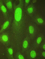
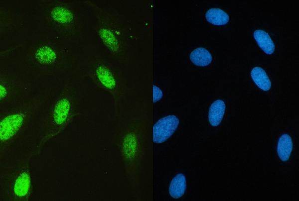
ARG59189 anti-USP7 / HAUSP antibody ICC/IF image
Immunofluorescence: U2OS cells were blocked with 10% goat serum and then stained with ARG59189 anti-USP7 / HAUSP antibody (green) at 2 µg/ml dilution, overnight at 4°C.
-
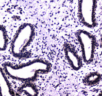
ARG59189 anti-USP7 / HAUSP antibody IHC-P image
Immunohistochemistry: Paraffin-embedded Human mammary cancer tissue. Antigen Retrieval: Heat mediated was performed in Citrate buffer (pH 6.0, epitope retrieval solution) for 20 min. The tissue section was blocked with 10% goat serum. The tissue section was then stained with ARG59189 anti-USP7 / HAUSP antibody at 1 µg/ml dilution, overnight at 4°C.
-
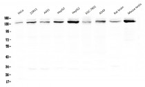
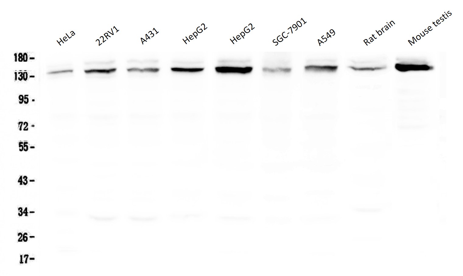
ARG59189 anti-USP7 / HAUSP antibody WB image
Western blot: 50 µg of samples under reducing conditions. HeLa, 22RV1, A431, HepG2, HepG2, SGC-7901, A549, Rat brain and Mouse testis lysates stained with ARG59189 anti-USP7 / HAUSP antibody at 0.5 µg/ml, overnight at 4°C.
-
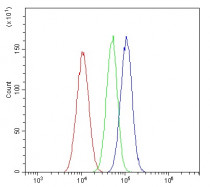
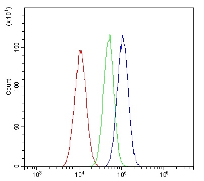
ARG59189 anti-USP7 / HAUSP antibody FACS image
Flow Cytometry: A549 cells were blocked with 10% normal goat serum and then stained with ARG59189 anti-USP7 / HAUSP antibody (blue) at 1 µg/10^6 cells for 30 min at 20°C, followed by incubation with DyLight®488 labelled secondary antibody. Isotype control antibody (green) was rabbit IgG (1 µg/10^6 cells) used under the same conditions. Unlabelled sample (red) was also used as a control.
-

ARG59189 anti-USP7 / HAUSP antibody ICC/IF image
Immunofluorescence: U2OS cells were blocked with 10% goat serum and then stained with ARG59189 anti-USP7 / HAUSP antibody (green) at 2 µg/ml dilution, overnight at 4°C. DAPI (blue) for nuclear staining.
-
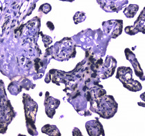
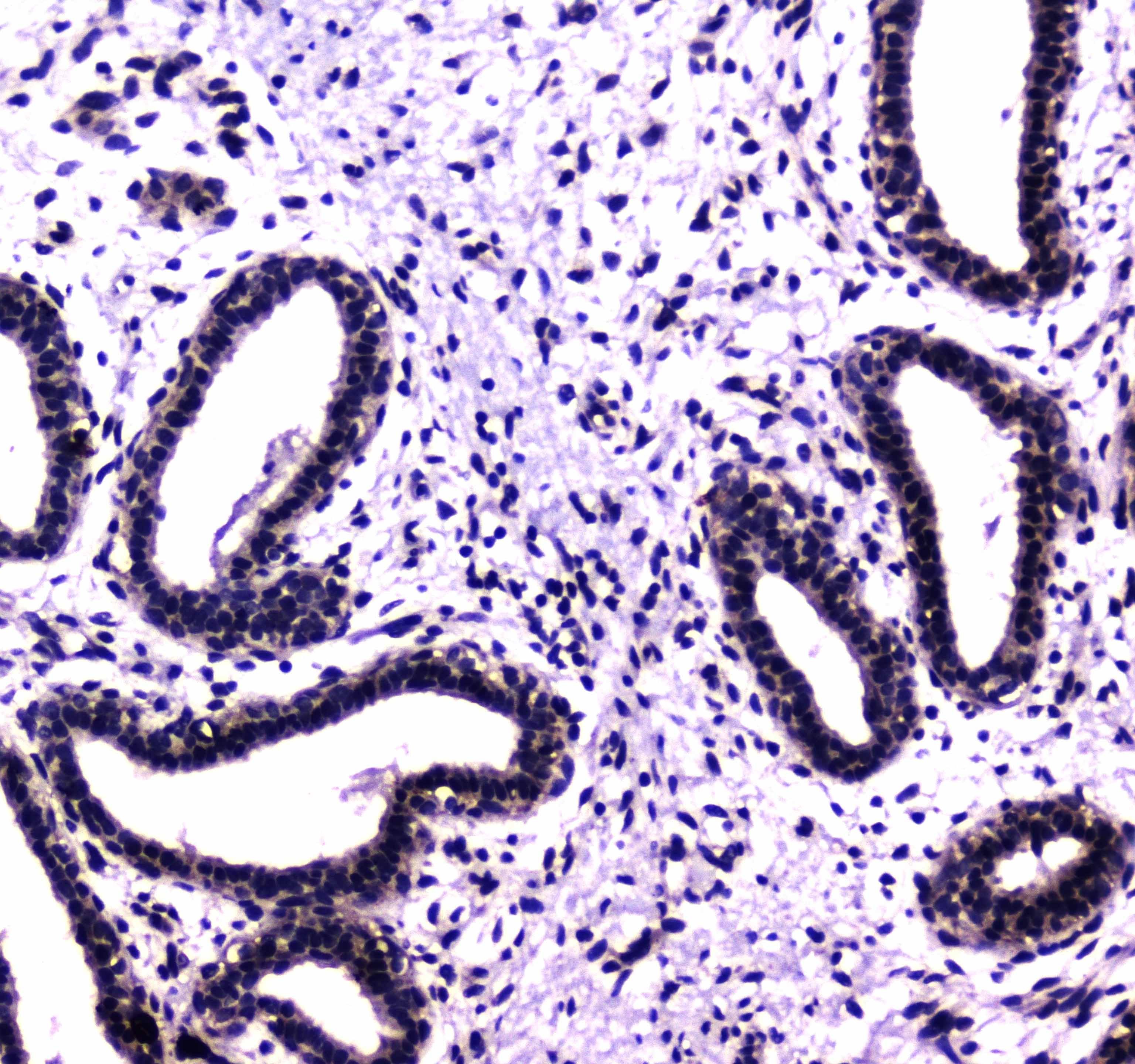
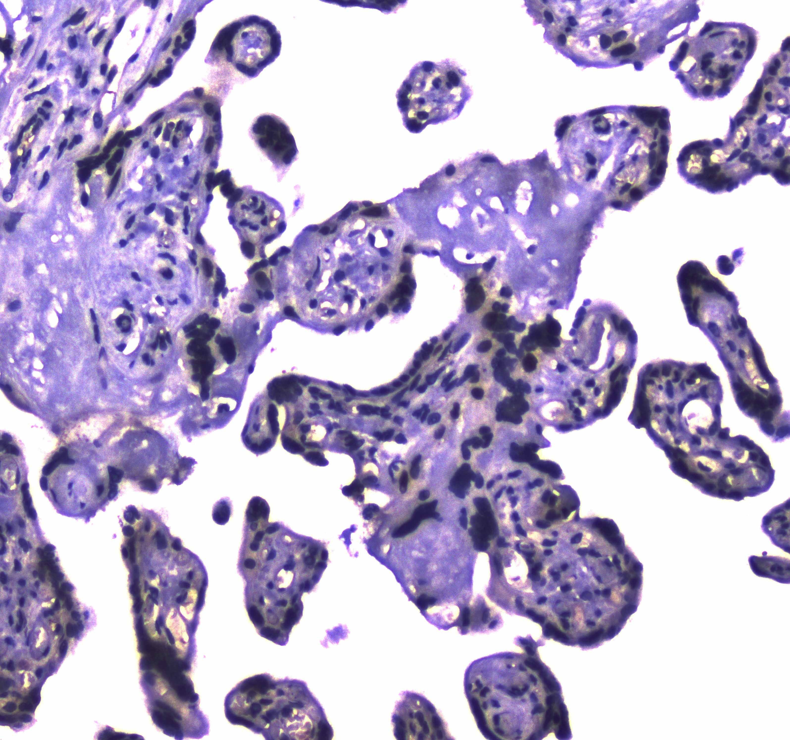
ARG59189 anti-USP7 / HAUSP antibody IHC-P image
Immunohistochemistry: Paraffin-embedded Human placenta tissue. Antigen Retrieval: Heat mediated was performed in Citrate buffer (pH 6.0, epitope retrieval solution) for 20 min. The tissue section was blocked with 10% goat serum. The tissue section was then stained with ARG59189 anti-USP7 / HAUSP antibody at 1 µg/ml dilution, overnight at 4°C.
-
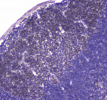
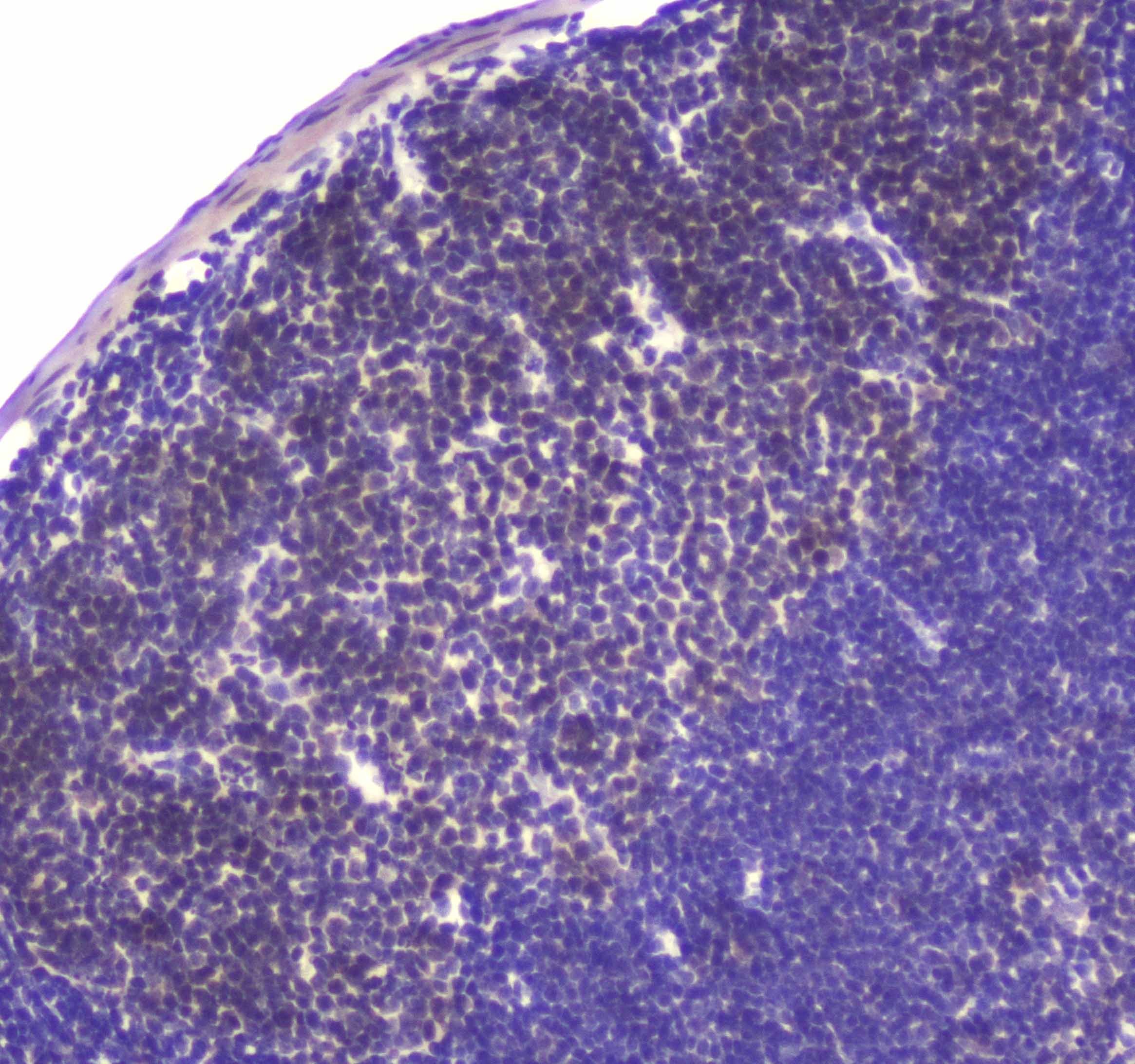
ARG59189 anti-USP7 / HAUSP antibody IHC-P image
Immunohistochemistry: Paraffin-embedded Mouse lymph nodes tissue. Antigen Retrieval: Heat mediated was performed in Citrate buffer (pH 6.0, epitope retrieval solution) for 20 min. The tissue section was blocked with 10% goat serum. The tissue section was then stained with ARG59189 anti-USP7 / HAUSP antibody at 1 µg/ml dilution, overnight at 4°C.