ARG52465
anti-UCHL1 / PGP9.5 antibody [BH7]
anti-UCHL1 / PGP9.5 antibody [BH7] for ICC/IF,IHC-Frozen sections,Western blot and Human,Mouse,Rat,Bovine,Pig
Cell Biology and Cellular Response antibody; Gene Regulation antibody; Neuroscience antibody
Overview
| Product Description | Mouse Monoclonal antibody [BH7] recognizes UCHL1 / PGP9.5 |
|---|---|
| Tested Reactivity | Hu, Ms, Rat, Bov, Pig |
| Predict Reactivity | Mamm |
| Tested Application | ICC/IF, IHC-Fr, WB |
| Host | Mouse |
| Clonality | Monoclonal |
| Clone | BH7 |
| Isotype | IgG1 |
| Target Name | UCHL1 / PGP9.5 |
| Antigen Species | Human |
| Immunogen | Recombinant full length human UCHL1 purified from E. coli |
| Conjugation | Un-conjugated |
| Alternate Names | PGP95; UCH-L1; PGP9.5; PARK5; Ubiquitin thioesterase L1; HEL-117; Neuron cytoplasmic protein 9.5; Uch-L1; EC 6.-.-.-; PGP 9.5; Ubiquitin carboxyl-terminal hydrolase isozyme L1; NDGOA; EC 3.4.19.12 |
Application Instructions
| Application Suggestion |
|
||||||||
|---|---|---|---|---|---|---|---|---|---|
| Application Note | Specific for the ~24kDa UCHL1 protein. * The dilutions indicate recommended starting dilutions and the optimal dilutions or concentrations should be determined by the scientist. |
Properties
| Form | Liquid |
|---|---|
| Purification | Total IgG fraction |
| Buffer | Total IgG fraction and 10 mM Sodium azide |
| Preservative | 10 mM Sodium azide |
| Storage Instruction | For continuous use, store undiluted antibody at 2-8°C for up to a week. For long-term storage, aliquot and store at -20°C or below. Storage in frost free freezers is not recommended. Avoid repeated freeze/thaw cycles. Suggest spin the vial prior to opening. The antibody solution should be gently mixed before use. |
| Note | For laboratory research only, not for drug, diagnostic or other use. |
Bioinformation
| Database Links | |
|---|---|
| Gene Symbol | UCHL1 |
| Gene Full Name | ubiquitin carboxyl-terminal esterase L1 (ubiquitin thiolesterase) |
| Background | Ubiquitin C-terminal hydrolase 1 (UCHL1) is also known as ubiquitin carboxyl esterase L1, ubiquitin thiolesterase, neuron-specific protein PGP9.5 and Park5. It was originally identified as a major component of the neuronal cytoplasm from 2-dimensional gel analysis of brain tissues, and was given the name PGP9.5 . It was later found that ubiquitin C-terminal hydrolase enzyme activity was associated with the PGP9.5 protein . The ubiquitin C-terminal hydrolases cleave ubiquitin from other molecules. Regulation of the ubiquitin pathway is very important and many disease states are associated with defects in this pathway. Genetic knockout of UCHL1 in mice results in a motor neuron degeneration similar to the spontaneous gracile axonal dystrophy (gad) mutant mice . Point mutations in the UCHL1 gene are associated with some forms of human Parkinson's disease . Since UCHL1 is heavily expressed in neurons, it is released in large amounts following injury or degeneration, so the detection of UCHL1 in CSF and other bodily fluids can be used as a biomarker. |
| Research Area | Cell Biology and Cellular Response antibody; Gene Regulation antibody; Neuroscience antibody |
| Calculated MW | 25 kDa |
| PTM | O-glycosylated. |
Images (5) Click the Picture to Zoom In
-
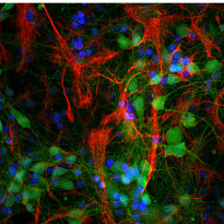
ARG52465 anti-UCHL1 / PGP9.5 antibody [BH7] ICC/IF image
Immunofluorescence: Cortical neuron-glial culture from E20 Rat stained with ARG52465 anti-UCHL1 / PGP9.5 antibody [BH7] (green) at 1:5000 dilution, and costained with anti-GFAP antibody (red) at 1:5000 dilution. DAPI (blue) for nuclear staining.
Clone BH7 stains cell bodies and dendrites of neurons, while the GFAP antibody labels astrocytes.
-
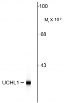
ARG52465 anti-UCHL1 / PGP9.5 antibody [BH7] WB image
Western blot: Rat hippocampal homogenate showing specific immunolabeling of the ~ 24k UCHL1 protein stained with ARG52465 anti-UCHL1 / PGP9.5 antibody [BH7].
-
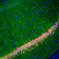
ARG52465 anti-UCHL1 / PGP9.5 antibody [BH7] IHC-Fr image
Immunohistochemistry: Frozen section of Rat hippocampal tissue stained with ARG52465 anti-UCHL1 / PGP9.5 antibody [BH7] (green) at 1:5000 dilution, and costained with ARG10712 anti-FOX3 / NeuN antibody (red) at 1:2000 dilution. DAPI (blue) for nuclear staining. Following transcardial perfusion of Rat with 4% paraformaldehyde, brain was post fixed for 24 hours, cut to 45 µM, and free-floating sections were stained with above antibodies.
The UCHL1 antibody stains the cell body and dendrites of hippocampal neurons, while the FOX3 antibody labels nuclei of the neuronal cells.
-
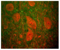
ARG52465 anti-UCHL1 / PGP9.5 antibody [BH7] IHC image
Immunohistochemistry: Rat spinal cord stained with ARG52465 anti-UCHL1 / PGP9.5 antibody [BH7] (red) and ARG52347 anti-Neurofilament NF-H antibody (green). The large cells are α-motorneurons and UCHL1 fills the cytoplasm of their perikarya and dendrites.
-
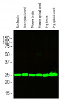
ARG52465 anti-UCHL1 / PGP9.5 antibody [BH7] WB image
Western blot: Rat brain, Rat spinal cord, Mouse brain, Mouse spinal cord, Pig brain and Pig spinal cord lysates stained with ARG52465 anti-UCHL1 / PGP9.5 antibody [BH7] (green) at 1:10000 dilution.


