ARG43060
anti-UBR2 antibody
anti-UBR2 antibody for Flow cytometry,ICC/IF,IHC-Formalin-fixed paraffin-embedded sections,Western blot and Human,Mouse
Overview
| Product Description | Rabbit Polyclonal antibody recognizes UBR2 |
|---|---|
| Tested Reactivity | Hu, Ms |
| Tested Application | FACS, ICC/IF, IHC-P, WB |
| Host | Rabbit |
| Clonality | Polyclonal |
| Isotype | IgG |
| Target Name | UBR2 |
| Antigen Species | Human |
| Immunogen | Recombinant protein corresponding to Q10-A664 of Human UBR2. |
| Conjugation | Un-conjugated |
| Alternate Names | C6orf133; bA49A4.1; EC 6.3.2.-; N-recognin-2; Ubiquitin-protein ligase E3-alpha-II; E3 ubiquitin-protein ligase UBR2; dJ392M17.3; Ubiquitin-protein ligase E3-alpha-2; dJ242G1.1 |
Application Instructions
| Application Suggestion |
|
||||||||||
|---|---|---|---|---|---|---|---|---|---|---|---|
| Application Note | IHC-P: Antigen Retrieval: Heat mediation was performed in EDTA buffer (pH 8.0). * The dilutions indicate recommended starting dilutions and the optimal dilutions or concentrations should be determined by the scientist. |
Properties
| Form | Liquid |
|---|---|
| Purification | Affinity purification with immunogen. |
| Buffer | 0.2% Na2HPO4, 0.9% NaCl, 0.05% Sodium azide and 4% Trehalose. |
| Preservative | 0.05% Sodium azide |
| Stabilizer | 4% Trehalose |
| Concentration | 0.5 mg/ml |
| Storage Instruction | For continuous use, store undiluted antibody at 2-8°C for up to a week. For long-term storage, aliquot and store at -20°C or below. Storage in frost free freezers is not recommended. Avoid repeated freeze/thaw cycles. Suggest spin the vial prior to opening. The antibody solution should be gently mixed before use. |
| Note | For laboratory research only, not for drug, diagnostic or other use. |
Bioinformation
| Database Links | |
|---|---|
| Gene Symbol | UBR2 |
| Gene Full Name | ubiquitin protein ligase E3 component n-recognin 2 |
| Background | This gene encodes an E3 ubiquitin ligase of the N-end rule proteolytic pathway that targets proteins with destabilizing N-terminal residues for polyubiquitylation and proteasome-mediated degradation. Alternative splicing results in multiple transcript variants. [provided by RefSeq, May 2010] |
| Function | E3 ubiquitin-protein ligase which is a component of the N-end rule pathway. Recognizes and binds to proteins bearing specific N-terminal residues that are destabilizing according to the N-end rule, leading to their ubiquitination and subsequent degradation. Plays a critical role in chromatin inactivation and chromosome-wide transcriptional silencing during meiosis via ubiquitination of histone H2A. Binds leucine and is a negative regulator of the leucine-mTOR signaling pathway, thereby controlling cell growth. Required for spermatogenesis, promotes, with Tex19.1, SPO11-dependent recombination foci to accumulate and drive robust homologous chromosome synapsis (By similarity). Polyubiquitinates LINE-1 retrotransposon encoded, LIRE1, which induces degradation, inhibiting LINE-1 retrotransposon mobilization (By similarity). [UniProt] |
| Cellular Localization | Nucleus. Note=Associated with chromatin during meiosis. [UniProt] |
| Calculated MW | 201 kDa |
Images (6) Click the Picture to Zoom In
-

ARG43060 anti-UBR2 antibody ICC/IF image
Immunofluorescence: HepG2 cells were blocked with 10% goat serum and then stained with ARG43060 anti-UBR2 antibody (green) at 2 µg/ml dilution, overnight at 4°C. DAPI (blue) for nuclear staining.
-

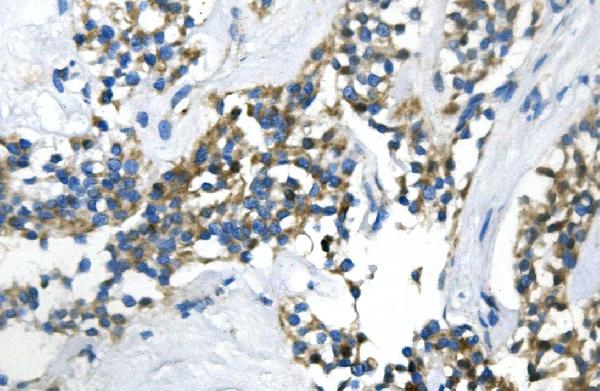
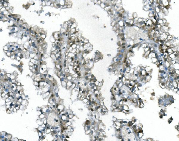
ARG43060 anti-UBR2 antibody IHC-P image
Immunohistochemistry: Paraffin-embedded Human pancreatic cancer tissue. Antigen Retrieval: Heat mediation was performed in EDTA buffer (pH 8.0). The tissue section was blocked with 10% goat serum. The tissue section was then stained with ARG43060 anti-UBR2 antibody at 1 µg/ml dilution, overnight at 4°C.
-
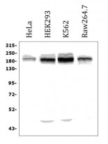
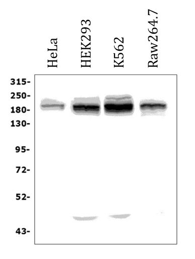
ARG43060 anti-UBR2 antibody WB image
Western blot: 50 µg of sample under reducing conditions. HeLa, HEK293, K562 and Raw264.7 cell lysates stained with ARG43060 anti-UBR2 antibody at 0.25 µg/ml dilution, overnight at 4°C.
-

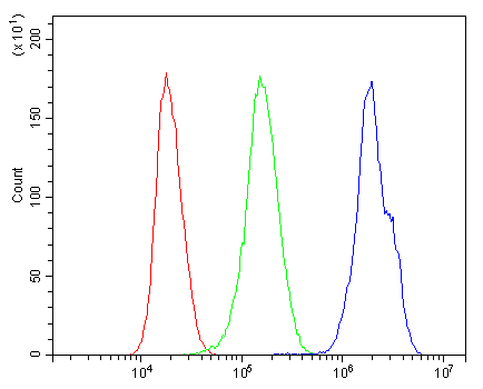
ARG43060 anti-UBR2 antibody FACS image
Flow Cytometry: HepG2 cells were blocked with 10% normal goat serum and then stained with ARG43060 anti-UBR2 antibody (blue) at 1 µg/10^6 cells for 30 min at 20°C, followed by incubation with DyLight®488 labelled secondary antibody. Isotype control antibody (green) was rabbit IgG (1 µg/10^6 cells) used under the same conditions. Unlabelled sample (red) was also used as a control.
-

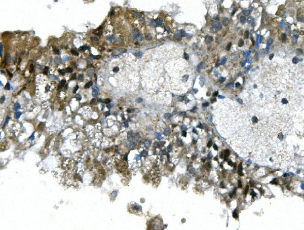
ARG43060 anti-UBR2 antibody IHC-P image
Immunohistochemistry: Paraffin-embedded Human renal cancer tissue. Antigen Retrieval: Heat mediation was performed in EDTA buffer (pH 8.0). The tissue section was blocked with 10% goat serum. The tissue section was then stained with ARG43060 anti-UBR2 antibody at 1 µg/ml dilution, overnight at 4°C.
-
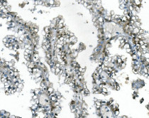
ARG43060 anti-UBR2 antibody IHC-P image
Immunohistochemistry: Paraffin-embedded Human renal cancer tissue. Antigen Retrieval: Heat mediation was performed in EDTA buffer (pH 8.0). The tissue section was blocked with 10% goat serum. The tissue section was then stained with ARG43060 anti-UBR2 antibody at 1 µg/ml dilution, overnight at 4°C.