ARG11149
anti-Tyrosine Hydroxylase antibody [4H2]
anti-Tyrosine Hydroxylase antibody [4H2] for ICC/IF,IHC-Frozen sections,Western blot and Human,Mouse,Rat
Overview
| Product Description | Mouse Monoclonal antibody [4H2] recognizes Tyrosine Hydroxylase |
|---|---|
| Tested Reactivity | Hu, Ms, Rat |
| Tested Application | ICC/IF, IHC-Fr, WB |
| Host | Mouse |
| Clonality | Monoclonal |
| Clone | 4H2 |
| Isotype | IgG1 |
| Target Name | Tyrosine Hydroxylase |
| Antigen Species | Human |
| Immunogen | Full-length Human Tyrosine Hydroxylase. |
| Conjugation | Un-conjugated |
| Alternate Names | DYT14; TYH; Tyrosine 3-monooxygenase; Tyrosine 3-hydroxylase; TH; DYT5b; EC 1.14.16.2 |
Application Instructions
| Application Suggestion |
|
||||||||
|---|---|---|---|---|---|---|---|---|---|
| Application Note | * The dilutions indicate recommended starting dilutions and the optimal dilutions or concentrations should be determined by the scientist. | ||||||||
| Observed Size | ~ 60 kDa |
Properties
| Form | Liquid |
|---|---|
| Purification | Purified |
| Buffer | PBS, 5 mM Sodium azide and 50% Glycerol. |
| Preservative | 5 mM Sodium azide |
| Stabilizer | 50% Glycerol |
| Concentration | 1 mg/ml |
| Storage Instruction | For continuous use, store undiluted antibody at 2-8°C for up to a week. For long-term storage, aliquot and store at -20°C. Storage in frost free freezers is not recommended. Avoid repeated freeze/thaw cycles. Suggest spin the vial prior to opening. The antibody solution should be gently mixed before use. |
| Note | For laboratory research only, not for drug, diagnostic or other use. |
Bioinformation
| Database Links | |
|---|---|
| Gene Symbol | TH |
| Gene Full Name | tyrosine hydroxylase |
| Function | Plays an important role in the physiology of adrenergic neurons. [UniProt] |
| Calculated MW | 59 kDa |
Images (2) Click the Picture to Zoom In
-
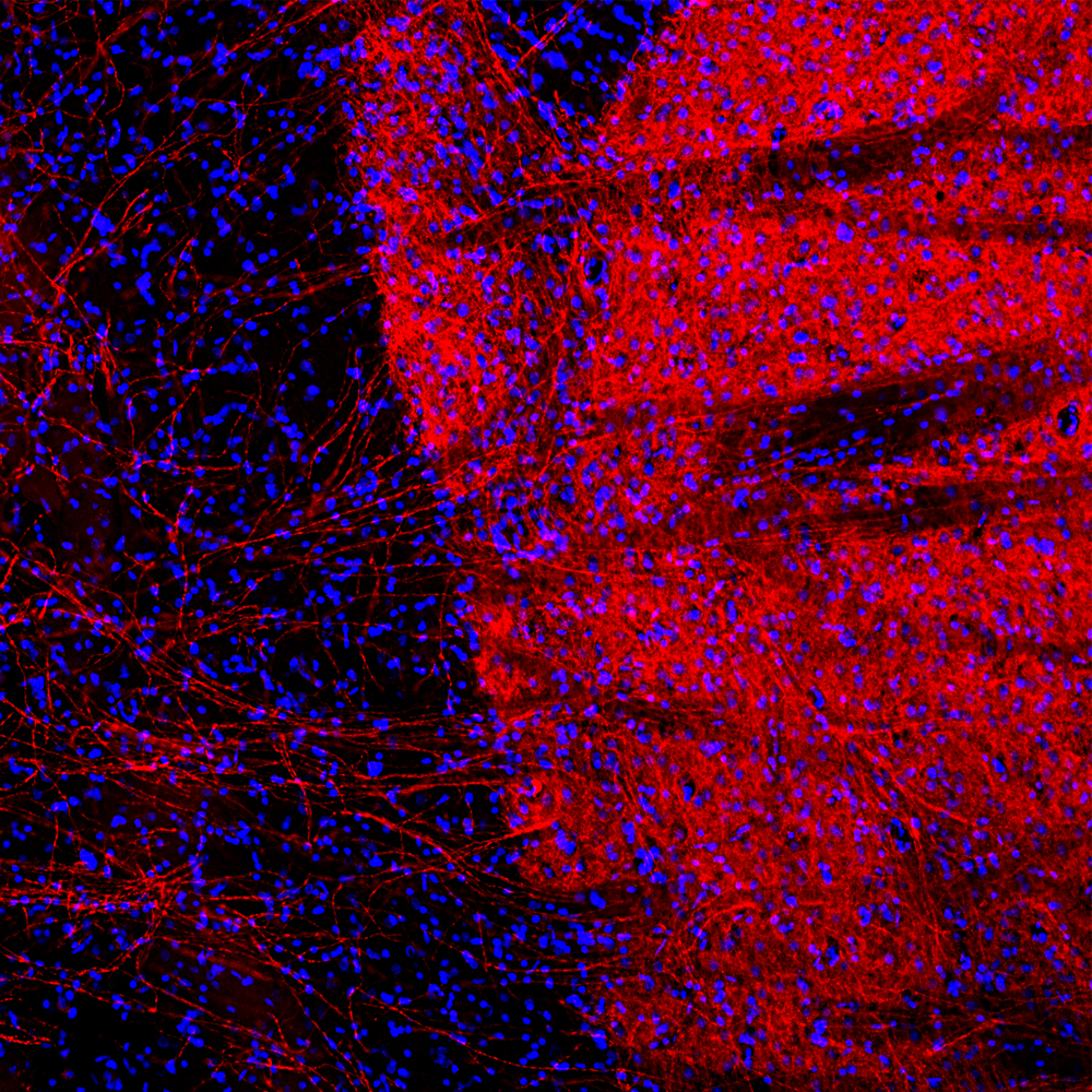
ARG11149 anti-Tyrosine Hydroxylase antibody [4H2] IHC-Fr image
Immunohistochemistry: Frozen section of Rat brain tissue stained with ARG11149 anti-Tyrosine Hydroxylase antibody [4H2] (red) at 1:1000 dilution. Hoechst (blue) for nuclear staining. (Sample preparation: Following transcardial perfusion of rat with 4% paraformaldehyde, brain was post fixed for 24 hours, cut to 45 µM, and free-floating sections were stained with the above antibodies.).
-

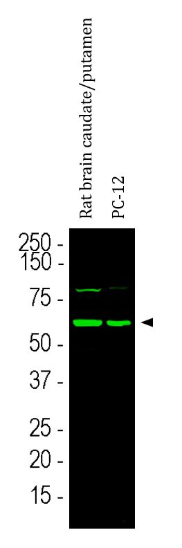
ARG11149 anti-Tyrosine Hydroxylase antibody [4H2] WB image
Western blot: Rat brain caudate/putamen and PC-12 cell lysates stained with ARG11149 anti-Tyrosine Hydroxylase antibody [4H2] at 1:5000 dilution.
The strong band at about 58 kDa corresponds to TH protein. The weak band at about 80 kDa in the rat brain homogenate is likely an aggregated form of TH, which does not affect the specific cell and process staining of this antibody.