ARG62652
anti-Tropomyosin antibody [TM31]
anti-Tropomyosin antibody [TM31] for IHC-Formalin-fixed paraffin-embedded sections,Western blot and Human,Mouse,Rat,Chicken,Rabbit
Cell Biology and Cellular Response antibody; Signaling Transduction antibody
Overview
| Product Description | Mouse Monoclonal antibody [TM-311] recognizes Tropomyosin |
|---|---|
| Tested Reactivity | Hu, Ms, Rat, Chk, Rb |
| Tested Application | IHC-P, WB |
| Host | Mouse |
| Clonality | Monoclonal |
| Clone | TM-311 |
| Isotype | IgG1, kappa |
| Target Name | Tropomyosin |
| Immunogen | Raised against purified gizzard Tropomyosin of chicken origin. |
| Conjugation | Un-conjugated |
| Alternate Names | C15orf13; Tropomyosin alpha-1 chain; TMSA; CMD1Y; HEL-S-265; HTM-alpha; CMH3; Tropomyosin-1; LVNC9; Alpha-tropomyosin |
Application Instructions
| Application Suggestion |
|
||||||
|---|---|---|---|---|---|---|---|
| Application Note | * The dilutions indicate recommended starting dilutions and the optimal dilutions or concentrations should be determined by the scientist. | ||||||
| Positive Control | LNCaP cells. Normal prostate or prostate carcinoma |
Properties
| Form | Liquid |
|---|---|
| Purification | Purified Antibody |
| Buffer | 1X PBS and 0.1% Sodium azide |
| Preservative | 0.1% Sodium azide |
| Concentration | 0.2 mg/ml |
| Storage Instruction | For continuous use, store undiluted antibody at 2-8°C for up to a week. For long-term storage, aliquot and store at -20°C or below. Storage in frost free freezers is not recommended. Avoid repeated freeze/thaw cycles. Suggest spin the vial prior to opening. The antibody solution should be gently mixed before use. |
| Note | For laboratory research only, not for drug, diagnostic or other use. |
Bioinformation
| Database Links | |
|---|---|
| Gene Symbol | TPM1 |
| Gene Full Name | tropomyosin 1 (alpha) |
| Background | This gene is a member of the tropomyosin family of highly conserved, widely distributed actin-binding proteins involved in the contractile system of striated and smooth muscles and the cytoskeleton of non-muscle cells. Tropomyosin is composed of two alpha-helical chains arranged as a coiled-coil. It is polymerized end to end along the two grooves of actin filaments and provides stability to the filaments. The encoded protein is one type of alpha helical chain that forms the predominant tropomyosin of striated muscle, where it also functions in association with the troponin complex to regulate the calcium-dependent interaction of actin and myosin during muscle contraction. In smooth muscle and non-muscle cells, alternatively spliced transcript variants encoding a range of isoforms have been described. Mutations in this gene are associated with type 3 familial hypertrophic cardiomyopathy. [provided by RefSeq, Jul 2008] |
| Function | Binds to actin filaments in muscle and non-muscle cells. Plays a central role, in association with the troponin complex, in the calcium dependent regulation of vertebrate striated muscle contraction. Smooth muscle contraction is regulated by interaction with caldesmon. In non-muscle cells is implicated in stabilizing cytoskeleton actin filaments. [UniProt] |
| Research Area | Cell Biology and Cellular Response antibody; Signaling Transduction antibody |
| Calculated MW | 33 kDa |
| PTM | Phosphorylated at Ser-283 by DAPK1 in response to oxidative stress and this phosphorylation enhances stress fiber formation in endothelial cells. |
Images (1) Click the Picture to Zoom In
-
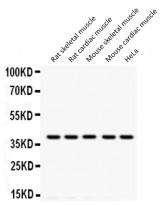
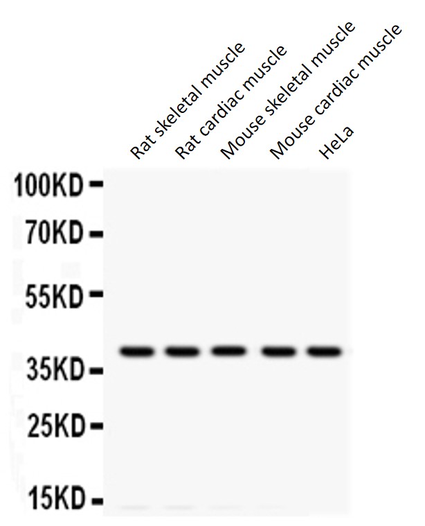
ARG62652 anti-Tropomyosin antibody [TM31] WB image
Western blot: Rat skeletal muscle, Rat cardiac muscle, Mouse skeletal muscle, Mouse cardiac muscle and HeLa whole cell lysates stained with ARG62652 anti-Tropomyosin antibody [TM31] at 0.5 µg/ml dilution.
Clone References
Induction of functional tissue-engineered skeletal muscle constructs by defined electrical stimulation.
WB / Mouse


Differential actin-regulatory activities of Tropomodulin1 and Tropomodulin3 with diverse tropomyosin and actin isoforms.
WB / Rabbit
Effect of the myosin light chain kinase inhibitor ML-7 on the proteome of hearts subjected to ischemia-reperfusion injury.
WB / Rat
Functional evaluation of artificial skeletal muscle tissue constructs fabricated by a magnetic force-based tissue engineering technique.
WB / Mouse
Novel method for measuring active tension generation by C2C12 myotube using UV-crosslinked collagen film.
WB / Mouse


