ARG55817
anti-Topoisomerase I antibody
anti-Topoisomerase I antibody for Flow cytometry,Western blot and Human,Rat
Overview
| Product Description | Mouse Monoclonal antibody recognizes Topoisomerase I |
|---|---|
| Tested Reactivity | Hu, Rat |
| Tested Application | FACS, WB |
| Host | Mouse |
| Clonality | Monoclonal |
| Clone | 1291CT875.142.166 |
| Isotype | IgG1, kappa |
| Target Name | Topoisomerase I |
| Antigen Species | Human |
| Immunogen | Recombinant protein (N-terminus) of Human Topoisomerase I |
| Conjugation | Un-conjugated |
| Protein Full Name | DNA topoisomerase 1 |
| Alternate Names | DNA topoisomerase 1; DNA topoisomerase I; TOPI; EC 5.99.1.2; Scl-70 Antigen; Topoisomerase (DNA) I |
Application Instructions
| Application Suggestion |
|
||||||
|---|---|---|---|---|---|---|---|
| Application Note | * The dilutions indicate recommended starting dilutions and the optimal dilutions or concentrations should be determined by the scientist. | ||||||
| Positive Control | MCF7 |
Properties
| Form | Liquid |
|---|---|
| Purification | Purification with Protein G. |
| Buffer | PBS and 0.09% (W/V) Sodium azide |
| Preservative | 0.09% (W/V) Sodium azide |
| Storage Instruction | For continuous use, store undiluted antibody at 2-8°C for up to a week. For long-term storage, aliquot and store at -20°C or below. Storage in frost free freezers is not recommended. Avoid repeated freeze/thaw cycles. Suggest spin the vial prior to opening. The antibody solution should be gently mixed before use. |
| Note | For laboratory research only, not for drug, diagnostic or other use. |
Bioinformation
| Database Links | |
|---|---|
| Gene Symbol | TOP1 |
| Gene Full Name | topoisomerase (DNA) I |
| Background | This gene encodes a DNA topoisomerase, an enzyme that controls and alters the topologic states of DNA during transcription. This enzyme catalyzes the transient breaking and rejoining of a single strand of DNA which allows the strands to pass through one another, thus altering the topology of DNA. This gene is localized to chromosome 20 and has pseudogenes which reside on chromosomes 1 and 22. [provided by RefSeq, Jul 2008] |
| Function | Releases the supercoiling and torsional tension of DNA introduced during the DNA replication and transcription by transiently cleaving and rejoining one strand of the DNA duplex. Introduces a single-strand break via transesterification at a target site in duplex DNA. The scissile phosphodiester is attacked by the catalytic tyrosine of the enzyme, resulting in the formation of a DNA-(3'-phosphotyrosyl)-enzyme intermediate and the expulsion of a 5'-OH DNA strand. The free DNA strand then undergoes passage around the unbroken strand thus removing DNA supercoils. Finally, in the religation step, the DNA 5'-OH attacks the covalent intermediate to expel the active-site tyrosine and restore the DNA phosphodiester backbone (By similarity). Regulates the alternative splicing of tissue factor (F3) pre-mRNA in endothelial cells. Involved in the circadian transcription of the core circadian clock component ARNTL/BMAL1 by altering the chromatin structure around the ROR response elements (ROREs) on the ARNTL/BMAL1 promoter. [UniProt] |
| Cellular Localization | Nucleus, nucleolus. Nucleus, nucleoplasm. Note=Diffuse nuclear localization with some enrichment in nucleoli. On CPT treatment, cleared from nucleoli into nucleoplasm. Sumolyated forms found in both nucleoplasm and nucleoli |
| Calculated MW | 91 kDa |
| PTM | Sumoylated. Lys-117 is the main site of sumoylation. Sumoylation plays a role in partitioning TOP1 between nucleoli and nucleoplasm. Levels are dramatically increased on camptothecin (CPT) treatment. Phosphorylation at Ser-506 by CK2 increases binding to supercoiled DNA and sensitivity to camptothecin. |
Images (2) Click the Picture to Zoom In
-

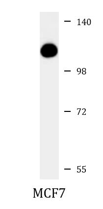
ARG55817 anti-Topoisomerase I antibody WB image
Western blot: 35 µg of MCF7 cell lysate stained with ARG55817 anti-Topoisomerase I antibody at 1:1000 dilution.
-
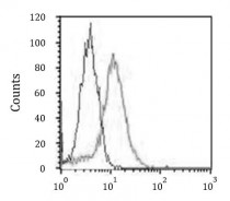
ARG55817 anti-Topoisomerase I antibody FACS image
Flow Cytometry: HeLa cells stained with ARG55817 anti-Topoisomerase I antibody (right histogram) at 1:25 dilution or isotype control antibody (left histogram), followed by incubation with Alexa Fluor® 488 labelled secondary antibody.