ARG52441
anti-Tau antibody
anti-Tau antibody for ICC/IF,Western blot and Human,Mouse,Rat
Neuroscience antibody; Signaling Transduction antibody; Neuron Development Study antibody
Overview
| Product Description | Chicken Polyclonal antibody recognizes Tau |
|---|---|
| Tested Reactivity | Hu, Ms, Rat |
| Tested Application | ICC/IF, WB |
| Host | Chicken |
| Clonality | Polyclonal |
| Isotype | IgY |
| Target Name | Tau |
| Antigen Species | Human |
| Immunogen | Recombinant full length 441 amino acid human tau isoform 2 (NP_005901.2) expressed in and purified from E.Coli. |
| Conjugation | Un-conjugated |
| Alternate Names | TAU; Neurofibrillary tangle protein; Paired helical filament-tau; PPND; DDPAC; FTDP-17; MTBT2; Microtubule-associated protein tau; PHF-tau; MSTD; PPP1R103; MTBT1; MAPTL |
Application Instructions
| Application Suggestion |
|
||||||
|---|---|---|---|---|---|---|---|
| Application Note | Specific for the ~48, 65 & 75k tau isoforms. * The dilutions indicate recommended starting dilutions and the optimal dilutions or concentrations should be determined by the scientist. |
Properties
| Form | Liquid |
|---|---|
| Purification | Total IgY fraction |
| Buffer | Total IgY fraction in PBS and 10 mM Sodium azide |
| Preservative | 10 mM Sodium azide |
| Storage Instruction | For continuous use, store undiluted antibody at 2-8°C for up to a week. For long-term storage, aliquot and store at -20°C or below. Storage in frost free freezers is not recommended. Avoid repeated freeze/thaw cycles. Suggest spin the vial prior to opening. The antibody solution should be gently mixed before use. |
| Note | For laboratory research only, not for drug, diagnostic or other use. |
Bioinformation
| Database Links | |
|---|---|
| Gene Symbol | MAPT |
| Gene Full Name | microtubule-associated protein tau |
| Background | Tau is a key microtubule-associated protein that plays an important role in the formation of microtubules in axons (Binder et al. 1985). Six tau isoforms have been identified as products of a single gene produced by alternative mRNA splicing (Goedert 1990). Tau mutations have been implicated in many neurodegenerative disorders such as Alzheimer’s disease (AD), Pick’s disease and progressive supranuclear palsy |
| Highlight | Related Antibody Duos and Panels: ARG30083 Neuron Development Marker Antibody Duo (Nestin, Tau) Related products: Tau antibodies; Tau ELISA Kits; Tau Duos / Panels; Anti-Chicken IgY secondary antibodies; Related news: 14-3-3η as a promising target for the treatment of Major Depression Disorder Neuronal Development Marker |
| Research Area | Neuroscience antibody; Signaling Transduction antibody; Neuron Development Study antibody |
| Calculated MW | 79 kDa |
| PTM | Phosphorylation at serine and threonine residues in S-P or T-P motifs by proline-directed protein kinases (PDPK1: CDK1, CDK5, GSK3, MAPK) (only 2-3 sites per protein in interphase, seven-fold increase in mitosis, and in the form associated with paired helical filaments (PHF-tau)), and at serine residues in K-X-G-S motifs by MAP/microtubule affinity-regulating kinase (MARK1 or MARK2), causing detachment from microtubules, and their disassembly. Phosphorylation decreases with age. Phosphorylation within tau/MAP's repeat domain or in flanking regions seems to reduce tau/MAP's interaction with, respectively, microtubules or plasma membrane components. Phosphorylation on Ser-610, Ser-622, Ser-641 and Ser-673 in several isoforms during mitosis. Phosphorylation at Ser-548 by GSK3B reduces ability to bind and stabilize microtubules. Phosphorylation at Ser-579 by BRSK1 and BRSK2 in neurons affects ability to bind microtubules and plays a role in neuron polarization. Phosphorylated at Ser-554, Ser-579, Ser-602, Ser-606 and Ser-669 by PHK. Phosphorylation at Ser-214 by SGK1 mediates microtubule depolymerization and neurite formation in hippocampal neurons. There is a reciprocal down-regulation of phosphorylation and O-GlcNAcylation. Phosphorylation on Ser-717 completely abolishes the O-GlcNAcylation on this site, while phosphorylation on Ser-713 and Ser-721 reduces glycosylation by a factor of 2 and 4 respectively. Phosphorylation on Ser-721 is reduced by about 41.5% by GlcNAcylation on Ser-717. Dephosphorylated at several serine and threonine residues by the serine/threonine phosphatase PPP5C. Polyubiquitinated. Requires functional TRAF6 and may provoke SQSTM1-dependent degradation by the proteasome (By similarity). PHF-tau can be modified by three different forms of polyubiquitination. 'Lys-48'-linked polyubiquitination is the major form, 'Lys-6'-linked and 'Lys-11'-linked polyubiquitination also occur. O-glycosylated. O-GlcNAcylation content is around 8.2%. There is reciprocal down-regulation of phosphorylation and O-GlcNAcylation. Phosphorylation on Ser-717 completely abolishes the O-GlcNAcylation on this site, while phosphorylation on Ser-713 and Ser-721 reduces O-GlcNAcylation by a factor of 2 and 4 respectively. O-GlcNAcylation on Ser-717 decreases the phosphorylation on Ser-721 by about 41.5%. Glycation of PHF-tau, but not normal brain TAU/MAPT. Glycation is a non-enzymatic post-translational modification that involves a covalent linkage between a sugar and an amino group of a protein molecule forming ketoamine. Subsequent oxidation, fragmentation and/or cross-linking of ketoamine leads to the production of advanced glycation endproducts (AGES). Glycation may play a role in stabilizing PHF aggregation leading to tangle formation in AD. |
Images (2) Click the Picture to Zoom In
-
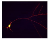
ARG52441 anti-Tau antibody ICC/IF image
Immunofluorescence: cultured rat hippocampal neurons stained with Tau antibody (ARG52441) showing staining of tau in red along the neuronal processes.
-
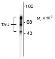
ARG52441 anti-Tau antibody WB image
Western Blot: rat cortex lysate stained with Tau antibody (ARG52441) showing specific immunolabeling of the ~48, 65 & 75k tau isoforms.


