ARG64874
anti-TSPO / PBR antibody
anti-TSPO / PBR antibody for Flow cytometry,ICC/IF,IHC-Formalin-fixed paraffin-embedded sections,Western blot and Mouse
Cell Biology and Cellular Response antibody; Cell Death antibody; Metabolism antibody; Signaling Transduction antibody
Overview
| Product Description | Goat Polyclonal antibody recognizes TSPO / PBR |
|---|---|
| Tested Reactivity | Ms |
| Predict Reactivity | Hu, Rat |
| Tested Application | FACS, ICC/IF, IHC-P, WB |
| Host | Goat |
| Clonality | Polyclonal |
| Isotype | IgG |
| Target Name | TSPO / PBR |
| Antigen Species | Mouse |
| Immunogen | C-RDNSGRRGGSRLPE |
| Conjugation | Un-conjugated |
| Alternate Names | DBI; BZRP; PBS; PBR; pk18; PKBS; mDRC; BPBS; IBP; Translocator protein; Mitochondrial benzodiazepine receptor; PTBR; MBR; Peripheral-type benzodiazepine receptor |
Application Instructions
| Application Suggestion |
|
||||||||||
|---|---|---|---|---|---|---|---|---|---|---|---|
| Application Note | WB: Recommend incubate at RT for 1h. IHC-P: Antigen Retrieval: Heat mediation was performed in Citrate buffer (pH 6.0). * The dilutions indicate recommended starting dilutions and the optimal dilutions or concentrations should be determined by the scientist. |
Properties
| Form | Liquid |
|---|---|
| Purification | Purified from goat serum by ammonium sulphate precipitation followed by antigen affinity chromatography using the immunizing peptide. |
| Buffer | Tris saline (pH 7.3), 0.02% Sodium azide and 0.5% BSA |
| Preservative | 0.02% Sodium azide |
| Stabilizer | 0.5% BSA |
| Concentration | 0.5 mg/ml |
| Storage Instruction | For continuous use, store undiluted antibody at 2-8°C for up to a week. For long-term storage, aliquot and store at -20°C or below. Storage in frost free freezers is not recommended. Avoid repeated freeze/thaw cycles. Suggest spin the vial prior to opening. The antibody solution should be gently mixed before use. |
| Note | For laboratory research only, not for drug, diagnostic or other use. |
Bioinformation
| Database Links | |
|---|---|
| Gene Symbol | Tspo |
| Gene Full Name | translocator protein |
| Background | Present mainly in the mitochondrial compartment of peripheral tissues, the protein encoded by this gene interacts with some benzodiazepines and has different affinities than its endogenous counterpart. The protein is a key factor in the flow of cholesterol into mitochondria to permit the initiation of steroid hormone synthesis. Alternatively spliced transcript variants have been reported; one of the variants lacks an internal exon and is considered non-coding, and the other variants encode the same protein. [provided by RefSeq, Feb 2012] |
| Function | Can bind protoporphyrin IX and may play a role in the transport of porphyrins and heme (By similarity). Was initially identified as peripheral-type benzodiazepine receptor; can also bind isoquinoline carboxamides. Promotes the transport of cholesterol across mitochondrial membranes and may play a role in lipid metabolism (PubMed:9832438, PubMed:24814875), but its precise physiological role is controversial. According to some reports, it is not required for steroid hormone biosynthesis (PubMed:24174323, PubMed:24936060). [UniProt] |
| Research Area | Cell Biology and Cellular Response antibody; Cell Death antibody; Metabolism antibody; Signaling Transduction antibody |
| Calculated MW | 19 kDa |
Images (6) Click the Picture to Zoom In
-
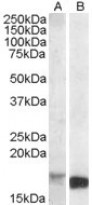
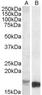
ARG64874 anti-TSPO / PBR antibody WB image
Western blot: Mouse Kidney lysate (A) and Mouse Spleen lysate (B) (35 µg protein in RIPA buffer). stained with ARG64874 anti-TSPO / PBR antibody at 1 µg/ml dilution.
-
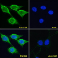
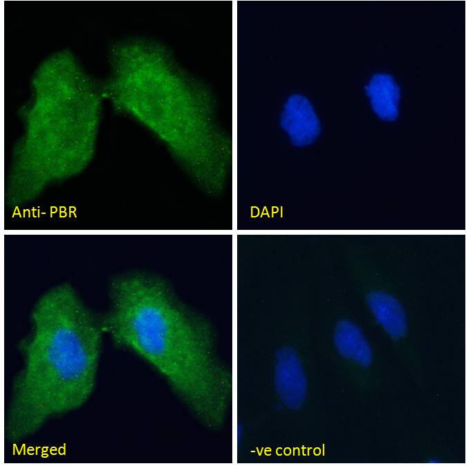
ARG64874 anti-TSPO / PBR antibody ICC/IF image
Immunofluorescence: Paraformaldehyde fixed NIH/3T3 cells permeabilized with 0.15% Triton. Cells were stained with ARG64874 anti-TSPO / PBR antibody (green) at 10 µg/ml dilution for 1 hour. DAPI (blue) for nuclear staining. Negative control: Unimmunized goat IgG (green) at 10 µg/ml dilution.
-

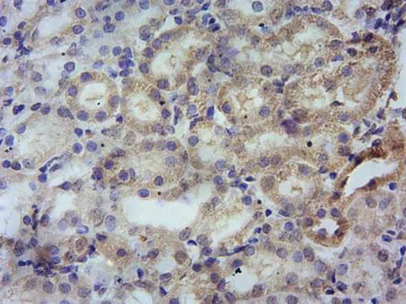
ARG64874 anti-TSPO / PBR antibody IHC-P image
Immunohistochemistry: Paraffin-embedded Mouse kidney tissue. Antigen Retrieval: Heat mediation was performed in Citrate buffer (pH 6.0). The tissue section was stained with ARG64874 anti-TSPO / PBR antibody at 10 µg/ml dilution followed by HRP-staining.
-
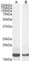
ARG64874 anti-TSPO / PBR antibody WB image
Western blot: 35 µg of Mouse kidney (A) and NIH/3T3 (B) cell lysates (in RIPA buffer) stained with ARG64874 anti-TSPO / PBR antibody at 0.03 µg/ml (A) and 0.01 µg/ml (B) dilutions and incubated at RT for 1 hour.
-

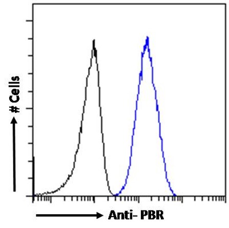
ARG64874 anti-TSPO / PBR antibody FACS image
Flow Cytometry: Paraformaldehyde-fixed NIH/3T3 cells permeabilized with 0.5% Triton. Cells were stained with ARG64874 anti-TSPO / PBR antibody (blue line) at 10 µg/ml dilution for 1 hour, followed by incubation with Alexa FluorR 488 labelled secondary antibody. IgG control: Unimmunized goat IgG (black line).
-

ARG64874 anti-TSPO / PBR antibody ICC/IF image
Immunofluorescence: Paraformaldehyde fixed HeLa cells permeabilized with 0.15% Triton. Cells were stained with ARG64874 anti-TSPO / PBR antibody (green) at 10 µg/ml dilution for 1 hour. DAPI (blue) for nuclear staining. Negative control: Unimmunized goat IgG (green) at 10 µg/ml dilution.