ARG53674
anti-TNFR2 antibody
anti-TNFR2 antibody for IHC-Formalin-fixed paraffin-embedded sections and Human
Cancer antibody; Cell Biology and Cellular Response antibody; Cell Death antibody; Immune System antibody; Neuroscience antibody; Signaling Transduction antibody
Overview
| Product Description | Rabbit Polyclonal antibody recognizes TNFR2 |
|---|---|
| Tested Reactivity | Hu |
| Tested Application | IHC-P |
| Host | Rabbit |
| Clonality | Polyclonal |
| Isotype | IgG |
| Target Name | TNFR2 |
| Antigen Species | Mouse |
| Immunogen | Synthetic peptide derived from C-terminus of mouse TNF-R2. |
| Conjugation | Un-conjugated |
| Alternate Names | TNFR1B; CD antigen CD120b; p75; CD120b; p75TNFR; TNF-R-II; TNFBR; TBPII; TNFR80; TBP-2; TNFR2; Tumor necrosis factor receptor 2; Tumor necrosis factor receptor type II; Etanercept; Tumor necrosis factor receptor superfamily member 1B; TNFR-II; TNF-R75; TNF-R2; TNF-RII; p80 TNF-alpha receptor |
Application Instructions
| Application Suggestion |
|
||||
|---|---|---|---|---|---|
| Application Note | IHC-P: Antigen Retrieval: Boil tissue section in 10mM citrate buffer, pH 6.0 for 10 min followed by cooling at RT for 20 min. Incubation Time: 30 min at RT. * The dilutions indicate recommended starting dilutions and the optimal dilutions or concentrations should be determined by the scientist. |
||||
| Positive Control | Liver Carcinoma, Thymus |
Properties
| Form | Liquid |
|---|---|
| Purification | Immunogen affinity purified |
| Buffer | PBS (pH 7.6), 1% BSA and < 0.1% Sodium azide |
| Preservative | < 0.1% Sodium azide |
| Stabilizer | 1% BSA |
| Storage Instruction | For continuous use, store undiluted antibody at 2-8°C for up to a week. For long-term storage, aliquot and store at -20°C or below. Storage in frost free freezers is not recommended. Avoid repeated freeze/thaw cycles. Suggest spin the vial prior to opening. The antibody solution should be gently mixed before use. |
| Note | For laboratory research only, not for drug, diagnostic or other use. |
Bioinformation
| Database Links |
Swiss-port # P20333 Human Tumor necrosis factor receptor superfamily member 1B |
|---|---|
| Gene Symbol | Tnfrsf1b |
| Gene Full Name | tumor necrosis factor receptor superfamily, member 1b |
| Background | The protein encoded by this gene is a member of the TNF-receptor superfamily. This protein and TNF-receptor 1 form a heterocomplex that mediates the recruitment of two anti-apoptotic proteins, c-IAP1 and c-IAP2, which possess E3 ubiquitin ligase activity. The function of IAPs in TNF-receptor signalling is unknown, however, c-IAP1 is thought to potentiate TNF-induced apoptosis by the ubiquitination and degradation of TNF-receptor-associated factor 2, which mediates anti-apoptotic signals. Knockout studies in mice also suggest a role of this protein in protecting neurons from apoptosis by stimulating antioxidative pathways. [provided by RefSeq, Jul 2008] |
| Function | Receptor with high affinity for TNFSF2/TNF-alpha and approximately 5-fold lower affinity for homotrimeric TNFSF1/lymphotoxin-alpha. The TRAF1/TRAF2 complex recruits the apoptotic suppressors BIRC2 and BIRC3 to TNFRSF1B/TNFR2 (By similarity). [UniProt] |
| Cellular Localization | Cytoplasm |
| Research Area | Cancer antibody; Cell Biology and Cellular Response antibody; Cell Death antibody; Immune System antibody; Neuroscience antibody; Signaling Transduction antibody |
| Calculated MW | 48 kDa |
| PTM | Phosphorylated; mainly on serine residues and with a very low level on threonine residues. A soluble form (tumor necrosis factor binding protein 2) is produced from the membrane form by proteolytic processing. |
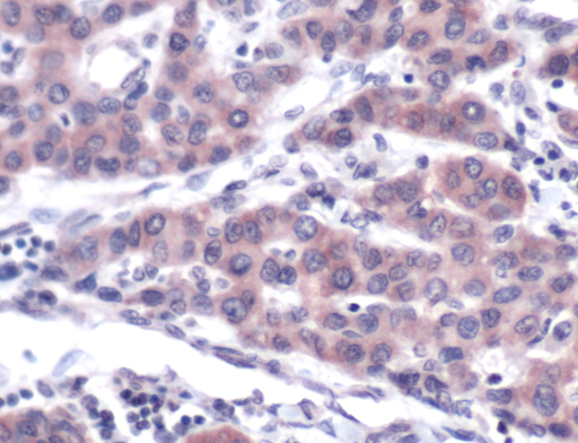
Images (1) Click the Picture to Zoom In
-
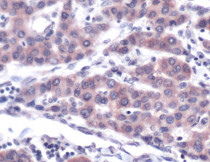
ARG53674 anti-TNFR2 antibody IHC-P image
Immunohistochemistry: Human Liver Carcinoma stained with ARG53674 anti-TNFR2 antibody.