ARG54267
anti-TNF alpha antibody [MAb11] (PE)
anti-TNF alpha antibody [MAb11] (PE) for Flow cytometry and Human,Primates,Pig
Cancer antibody; Cell Biology and Cellular Response antibody; Immune System antibody; Metabolism antibody; Signaling Transduction antibody
Overview
| Product Description | PE-conjugated Mouse Monoclonal antibody [MAb11] recognizes TNF-alpha |
|---|---|
| Tested Reactivity | Hu, NHuPrm, Pig |
| Tested Application | FACS |
| Specificity | The clone MAb11 recognizes human 17-26 kDa cytokine TNF-alpha (tumor necrosis factor alpha). |
| Host | Mouse |
| Clonality | Monoclonal |
| Clone | MAb11 |
| Isotype | IgG1 |
| Target Name | TNF alpha |
| Antigen Species | Human |
| Immunogen | Recombinant human TNF-alpha |
| Conjugation | PE |
| Alternate Names | Tumor necrosis factor ligand superfamily member 2; DIF; Cachectin; ICD2; ICD1; N-terminal fragment; TNF-a; TNFA; TNFSF2; TNF-alpha; Tumor necrosis factor; NTF |
Application Instructions
| Application Suggestion |
|
||||
|---|---|---|---|---|---|
| Application Note | * The dilutions indicate recommended starting dilutions and the optimal dilutions or concentrations should be determined by the scientist. |
Properties
| Form | Liquid |
|---|---|
| Purification Note | The purified antibody is conjugated with R-Phycoerythrin (PE) under optimum conditions. The conjugate is purified by size-exclusion chromatography and adjusted for direct use. No reconstitution is necessary. |
| Buffer | PBS, 15 mM Sodium azide and 0.2% (w/v) high-grade protease free BSA |
| Preservative | 15 mM Sodium azide |
| Stabilizer | 0.2% (w/v) high-grade protease free BSA |
| Storage Instruction | Aliquot and store in the dark at 2-8°C. Keep protected from prolonged exposure to light. Avoid repeated freeze/thaw cycles. Suggest spin the vial prior to opening. The antibody solution should be gently mixed before use. |
| Note | For laboratory research only, not for drug, diagnostic or other use. |
Bioinformation
| Database Links | |
|---|---|
| Gene Symbol | TNF |
| Gene Full Name | tumor necrosis factor |
| Background | TNF-alpha is a cytokine produced by monocytes, macrophages, neutrophils, NK cells, CD4+ T cells and many transformed cells. It can be expressed as a 17 kDa free molecule, or as a 26 kDa membrane protein. TNF-alpha easily forms stable trimers, but also other multimeric complexes. In the immune system, it is an important regulator, which has cytolytic and cytostatic activity against a range of tumor cells, increases fibroblast proliferation and supports neutrophil chemotaxis and phagocytosis. |
| Function | Cytokine that binds to TNFRSF1A/TNFR1 and TNFRSF1B/TNFBR. It is mainly secreted by macrophages and can induce cell death of certain tumor cell lines. It is potent pyrogen causing fever by direct action or by stimulation of interleukin-1 secretion and is implicated in the induction of cachexia, Under certain conditions it can stimulate cell proliferation and induce cell differentiation. Impairs regulatory T-cells (Treg) function in individuals with rheumatoid arthritis via FOXP3 dephosphorylation. Upregulates the expression of protein phosphatase 1 (PP1), which dephosphorylates the key 'Ser-418' residue of FOXP3, thereby inactivating FOXP3 and rendering Treg cells functionally defective (PubMed:23396208). The TNF intracellular domain (ICD) form induces IL12 production in dendritic cells. [UniProt] |
| Highlight | Related products: TNF alpha antibodies; TNF alpha ELISA Kits; TNF alpha Duos / Panels; TNF alpha recombinant proteins; Anti-Mouse IgG secondary antibodies; Related news: HMGB1 in inflammation Inflammatory Cytokines |
| Research Area | Cancer antibody; Cell Biology and Cellular Response antibody; Immune System antibody; Metabolism antibody; Signaling Transduction antibody |
| Calculated MW | 26 kDa |
| PTM | The soluble form derives from the membrane form by proteolytic processing. The membrane-bound form is further proteolytically processed by SPPL2A or SPPL2B through regulated intramembrane proteolysis producing TNF intracellular domains (ICD1 and ICD2) released in the cytosol and TNF C-domain 1 and C-domain 2 secreted into the extracellular space. The membrane form, but not the soluble form, is phosphorylated on serine residues. Dephosphorylation of the membrane form occurs by binding to soluble TNFRSF1A/TNFR1. O-glycosylated; glycans contain galactose, N-acetylgalactosamine and N-acetylneuraminic acid. |
Images (1) Click the Picture to Zoom In
-
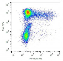
ARG54267 anti-TNF alpha antibody [MAb11] (PE) FACS image
Flow Cytometry: Activated human peripheral blood stained with ARG54267 anti-TNF alpha antibody [MAb11] (PE).


