ARG40571
anti-TNF alpha antibody (Biotin)
anti-TNF alpha antibody (Biotin) for ELISA,IHC-Formalin-fixed paraffin-embedded sections,Western blot and Human
Overview
| Product Description | Biotin-conjugated Rabbit Polyclonal antibody recognizes TNF alpha |
|---|---|
| Tested Reactivity | Hu |
| Tested Application | ELISA, IHC-P, WB |
| Host | Rabbit |
| Clonality | Polyclonal |
| Isotype | IgG |
| Target Name | TNF alpha |
| Antigen Species | Human |
| Immunogen | Human TNF alpha. |
| Conjugation | Biotin |
| Alternate Names | Tumor necrosis factor ligand superfamily member 2; DIF; Cachectin; ICD2; ICD1; N-terminal fragment; TNF-a; TNFA; TNFSF2; TNF-alpha; Tumor necrosis factor; NTF |
Application Instructions
| Application Suggestion |
|
||||||||
|---|---|---|---|---|---|---|---|---|---|
| Application Note | ELISA: ARG40571 (detection antibody) - ARG56622 (capture antibody) * The dilutions indicate recommended starting dilutions and the optimal dilutions or concentrations should be determined by the scientist. |
Properties
| Form | Liquid |
|---|---|
| Purification | Affinity purified. |
| Storage Instruction | Aliquot and store in the dark at 2-8°C. Keep protected from prolonged exposure to light. Avoid repeated freeze/thaw cycles. Suggest spin the vial prior to opening. The antibody solution should be gently mixed before use. |
| Note | For laboratory research only, not for drug, diagnostic or other use. |
Bioinformation
| Database Links | |
|---|---|
| Gene Symbol | TNF |
| Gene Full Name | tumor necrosis factor |
| Background | This gene encodes a multifunctional proinflammatory cytokine that belongs to the tumor necrosis factor (TNF) superfamily. This cytokine is mainly secreted by macrophages. It can bind to, and thus functions through its receptors TNFRSF1A/TNFR1 and TNFRSF1B/TNFBR. This cytokine is involved in the regulation of a wide spectrum of biological processes including cell proliferation, differentiation, apoptosis, lipid metabolism, and coagulation. This cytokine has been implicated in a variety of diseases, including autoimmune diseases, insulin resistance, and cancer. Knockout studies in mice also suggested the neuroprotective function of this cytokine. [provided by RefSeq, Jul 2008] |
| Function | Cytokine that binds to TNFRSF1A/TNFR1 and TNFRSF1B/TNFBR. It is mainly secreted by macrophages and can induce cell death of certain tumor cell lines. It is potent pyrogen causing fever by direct action or by stimulation of interleukin-1 secretion and is implicated in the induction of cachexia, Under certain conditions it can stimulate cell proliferation and induce cell differentiation. Impairs regulatory T-cells (Treg) function in individuals with rheumatoid arthritis via FOXP3 dephosphorylation. Upregulates the expression of protein phosphatase 1 (PP1), which dephosphorylates the key 'Ser-418' residue of FOXP3, thereby inactivating FOXP3 and rendering Treg cells functionally defective. Key mediator of cell death in the anticancer action of BCG-stimulated neutrophils in combination with DIABLO/SMAC mimetic in the RT4v6 bladder cancer cell line. The TNF intracellular domain (ICD) form induces IL12 production in dendritic cells. [UniProt] |
| Cellular Localization | Cell membrane; Single-pass type II membrane protein. Tumor necrosis factor, membrane form: Membrane; Single-pass type II membrane protein. Tumor necrosis factor, soluble form: Secreted. C-domain 1: Secreted. C-domain 2: Secreted. [UniProt] |
| Highlight | Related products: TNF alpha antibodies; TNF alpha ELISA Kits; TNF alpha Duos / Panels; TNF alpha recombinant proteins; Anti-Rabbit IgG secondary antibodies; Related news: HMGB1 in inflammation Inflammatory Cytokines |
| Calculated MW | 26 kDa |
| PTM | The soluble form derives from the membrane form by proteolytic processing. The membrane-bound form is further proteolytically processed by SPPL2A or SPPL2B through regulated intramembrane proteolysis producing TNF intracellular domains (ICD1 and ICD2) released in the cytosol and TNF C-domain 1 and C-domain 2 secreted into the extracellular space. The membrane form, but not the soluble form, is phosphorylated on serine residues. Dephosphorylation of the membrane form occurs by binding to soluble TNFRSF1A/TNFR1. O-glycosylated; glycans contain galactose, N-acetylgalactosamine and N-acetylneuraminic acid. [UniProt] |
Images (3) Click the Picture to Zoom In
-
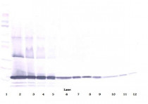
ARG40571 anti-TNF alpha antibody (Biotin) WB image
Western blot: 250 - 0.24 ng (left to right) of recombinant Human TNF alpha stained with ARG40571 anti-TNF alpha antibody (Biotin) at 0.1 - 0.2 µg/ml dilution, under reducing conditions.
-
ARG40571 anti-TNF alpha antibody (Biotin) ELISA image
ELISA: Human TNF alpha by sandwich ELISA (using 100 ul/well antibody solution), a concentration of 0.25 - 1.0 µg/ml of ARG40571 anti-TNF alpha antibody (Biotin) (detection antibody) in conjunction with ARG56622 anti-TNF alpha antibody (capture antibody), allows the detection of at least 0.2 - 0.4 ng/well of recombinant Human TNF alpha.
-
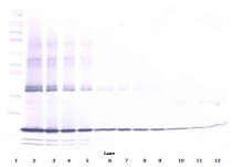
ARG40571 anti-TNF alpha antibody (Biotin) WB image
Western blot: 250 - 0.24 ng (left to right) of recombinant Human TNF alpha stained with ARG40571 anti-TNF alpha antibody (Biotin) at 0.1 - 0.2 µg/ml dilution, under non-reducing conditions.


