ARG42923
anti-TLR1 antibody
anti-TLR1 antibody for Flow cytometry,IHC-Formalin-fixed paraffin-embedded sections,Western blot and Human,Mouse,Rat
Overview
| Product Description | Rabbit Polyclonal antibody recognizes TLR1 |
|---|---|
| Tested Reactivity | Hu, Ms, Rat |
| Tested Application | FACS, IHC-P, WB |
| Host | Rabbit |
| Clonality | Polyclonal |
| Isotype | IgG |
| Target Name | TLR1 |
| Antigen Species | Human |
| Immunogen | Recombinant protein corresponding to F350-D404 of Human TLR1. |
| Conjugation | Un-conjugated |
| Alternate Names | CD281; CD antigen CD281; Toll/interleukin-1 receptor-like protein; TIL; Toll-like receptor 1; rsc786; TIL. LPRS5 |
Application Instructions
| Application Suggestion |
|
||||||||
|---|---|---|---|---|---|---|---|---|---|
| Application Note | IHC-P: Antigen Retrieval: Heat mediation was performed in Citrate buffer (pH 6.0) for 20 min. * The dilutions indicate recommended starting dilutions and the optimal dilutions or concentrations should be determined by the scientist. |
||||||||
| Observed Size | ~ 90 kDa |
Properties
| Form | Liquid |
|---|---|
| Purification | Affinity purification with immunogen. |
| Buffer | 0.2% Na2HPO4, 0.9% NaCl, 0.05% Sodium azide and 4% Trehalose. |
| Preservative | 0.05% Sodium azide |
| Stabilizer | 4% Trehalose |
| Concentration | 0.5 mg/ml |
| Storage Instruction | For continuous use, store undiluted antibody at 2-8°C for up to a week. For long-term storage, aliquot and store at -20°C or below. Storage in frost free freezers is not recommended. Avoid repeated freeze/thaw cycles. Suggest spin the vial prior to opening. The antibody solution should be gently mixed before use. |
| Note | For laboratory research only, not for drug, diagnostic or other use. |
Bioinformation
| Database Links | |
|---|---|
| Gene Symbol | TLR1 |
| Gene Full Name | toll-like receptor 1 |
| Background | The protein encoded by this gene is a member of the Toll-like receptor (TLR) family which plays a fundamental role in pathogen recognition and activation of innate immunity. TLRs are highly conserved from Drosophila to humans and share structural and functional similarities. They recognize pathogen-associated molecular patterns (PAMPs) that are expressed on infectious agents, and mediate the production of cytokines necessary for the development of effective immunity. The various TLRs exhibit different patterns of expression. This gene is ubiquitously expressed, and at higher levels than other TLR genes. Different length transcripts presumably resulting from use of alternative polyadenylation site, and/or from alternative splicing, have been noted for this gene. [provided by RefSeq, Jul 2008] |
| Function | Participates in the innate immune response to microbial agents. Specifically recognizes diacylated and triacylated lipopeptides. Cooperates with TLR2 to mediate the innate immune response to bacterial lipoproteins or lipopeptides (PubMed:21078852). Forms the activation cluster TLR2:TLR1:CD14 in response to triacylated lipopeptides, this cluster triggers signaling from the cell surface and subsequently is targeted to the Golgi in a lipid-raft dependent pathway (PubMed:16880211). Acts via MYD88 and TRAF6, leading to NF-kappa-B activation, cytokine secretion and the inflammatory response. [UniProt] |
| Cellular Localization | Cell membrane; Single-pass type I membrane protein. Cytoplasmic vesicle, phagosome membrane; Single-pass type I membrane protein. Membrane raft. Golgi apparatus. Note=Does not reside in lipid rafts before stimulation but accumulates increasingly in the raft upon the presence of the microbial ligand. In response to triacylated lipoproteins, TLR2:TLR1 heterodimers are recruited in lipid rafts, this recruitment determine the intracellular targeting to the Golgi apparatus. [UniProt] |
| Calculated MW | 90 kDa |
Images (6) Click the Picture to Zoom In
-
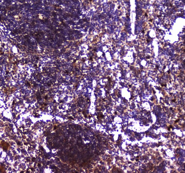
ARG42923 anti-TLR1 antibody IHC-P image
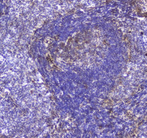
Immunohistochemistry: Paraffin-embedded Mouse spleen tissue. Antigen Retrieval: Heat mediation was performed in Citrate buffer (pH 6.0) for 20 min. The tissue section was blocked with 10% goat serum. The tissue section was then stained with ARG42923 anti-TLR1 antibody at 2 µg/ml dilution, overnight at 4°C.
-
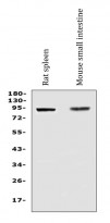
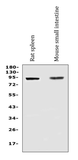
ARG42923 anti-TLR1 antibody WB image
Western blot: 50 µg of sample under reducing conditions. Rat spleen and Mouse small intestine lysates stained with ARG42923 anti-TLR1 antibody at 0.5 µg/ml dilution, overnight at 4°C.
-
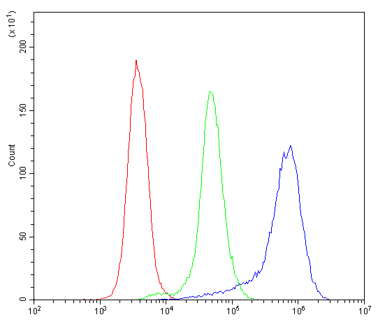
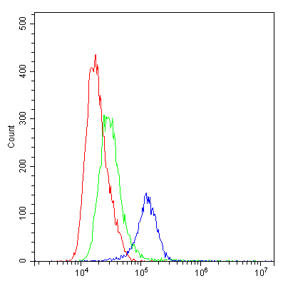
ARG42923 anti-TLR1 antibody FACS image
Flow Cytometry: K562 cells were blocked with 10% normal goat serum and then stained with ARG42923 anti-TLR1 antibody (blue) at 1 µg/10^6 cells for 30 min at 20°C, followed by incubation with DyLight®488 labelled secondary antibody. Isotype control antibody (green) was Rabbit IgG (1 µg/10^6 cells) used under the same conditions. Unlabelled sample (red) was also used as a control.
-
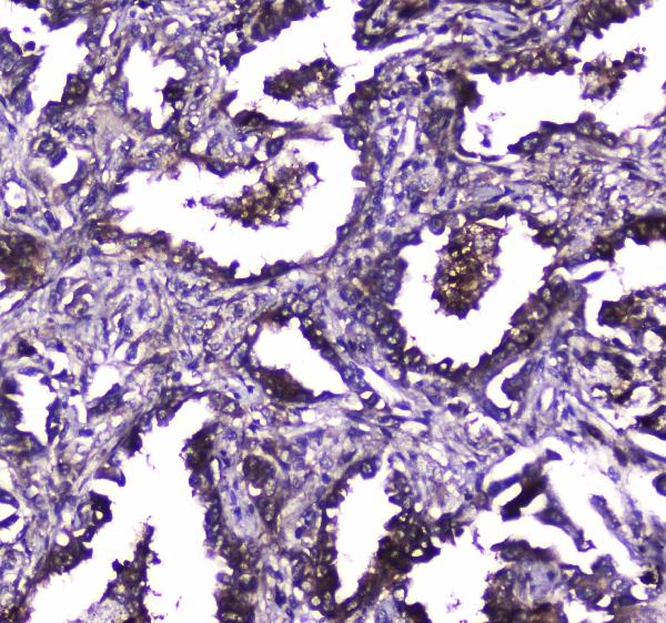
ARG42923 anti-TLR1 antibody IHC-P image
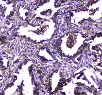
Immunohistochemistry: Paraffin-embedded Human lung cancer tissue. Antigen Retrieval: Heat mediation was performed in Citrate buffer (pH 6.0) for 20 min. The tissue section was blocked with 10% goat serum. The tissue section was then stained with ARG42923 anti-TLR1 antibody at 2 µg/ml dilution, overnight at 4°C.
-
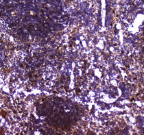
ARG42923 anti-TLR1 antibody IHC-P image
Immunohistochemistry: Paraffin-embedded Rat spleen tissue. Antigen Retrieval: Heat mediation was performed in Citrate buffer (pH 6.0) for 20 min. The tissue section was blocked with 10% goat serum. The tissue section was then stained with ARG42923 anti-TLR1 antibody at 2 µg/ml dilution, overnight at 4°C.
-
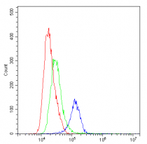
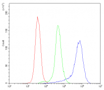
ARG42923 anti-TLR1 antibody FACS image
Flow Cytometry: THP-1 cells were blocked with 10% normal goat serum and then stained with ARG42923 anti-TLR1 antibody (blue) at 1 µg/10^6 cells for 30 min at 20°C, followed by incubation with DyLight®488 labelled secondary antibody. Isotype control antibody (green) was Rabbit IgG (1 µg/10^6 cells) used under the same conditions. Unlabelled sample (red) was also used as a control.