ARG55639
anti-TIMP1 antibody
anti-TIMP1 antibody for Flow cytometry,ICC/IF,IHC-Formalin-fixed paraffin-embedded sections,Western blot and Human
Overview
| Product Description | Rabbit Polyclonal antibody recognizes TIMP1 |
|---|---|
| Tested Reactivity | Hu |
| Tested Application | FACS, ICC/IF, IHC-P, WB |
| Host | Rabbit |
| Clonality | Polyclonal |
| Isotype | IgG |
| Target Name | TIMP1 |
| Antigen Species | Human |
| Immunogen | KLH-conjugated synthetic peptide corresponding to aa. 41-70 of Human TIMP1. |
| Conjugation | Un-conjugated |
| Alternate Names | Erythroid-potentiating activity; TIMP; Collagenase inhibitor; Fibroblast collagenase inhibitor; TIMP-1; EPO; CLGI; Tissue inhibitor of metalloproteinases 1; HCI; Metalloproteinase inhibitor 1; EPA |
Application Instructions
| Application Suggestion |
|
||||||||||
|---|---|---|---|---|---|---|---|---|---|---|---|
| Application Note | * The dilutions indicate recommended starting dilutions and the optimal dilutions or concentrations should be determined by the scientist. | ||||||||||
| Positive Control | CEM |
Properties
| Form | Liquid |
|---|---|
| Purification | This antibody is prepared by Saturated Ammonium Sulfate (SAS) precipitation followed by dialysis against PBS. |
| Buffer | PBS and 0.09% (W/V) Sodium azide |
| Preservative | 0.09% (W/V) Sodium azide |
| Storage Instruction | For continuous use, store undiluted antibody at 2-8°C for up to a week. For long-term storage, aliquot and store at -20°C or below. Storage in frost free freezers is not recommended. Avoid repeated freeze/thaw cycles. Suggest spin the vial prior to opening. The antibody solution should be gently mixed before use. |
| Note | For laboratory research only, not for drug, diagnostic or other use. |
Bioinformation
| Database Links | |
|---|---|
| Gene Symbol | TIMP1 |
| Gene Full Name | TIMP metallopeptidase inhibitor 1 |
| Background | This gene belongs to the TIMP gene family. The proteins encoded by this gene family are natural inhibitors of the matrix metalloproteinases (MMPs), a group of peptidases involved in degradation of the extracellular matrix. In addition to its inhibitory role against most of the known MMPs, the encoded protein is able to promote cell proliferation in a wide range of cell types, and may also have an anti-apoptotic function. Transcription of this gene is highly inducible in response to many cytokines and hormones. In addition, the expression from some but not all inactive X chromosomes suggests that this gene inactivation is polymorphic in human females. This gene is located within intron 6 of the synapsin I gene and is transcribed in the opposite direction. [provided by RefSeq, Jul 2008] |
| Function | Metalloproteinase inhibitor that functions by forming one to one complexes with target metalloproteinases, such as collagenases, and irreversibly inactivates them by binding to their catalytic zinc cofactor. Acts on MMP1, MMP2, MMP3, MMP7, MMP8, MMP9, MMP10, MMP11, MMP12, MMP13 and MMP16. Does not act on MMP14. Also functions as a growth factor that regulates cell differentiation, migration and cell death and activates cellular signaling cascades via CD63 and ITGB1. Plays a role in integrin signaling. Mediates erythropoiesis in vitro; but, unlike IL3, it is species-specific, stimulating the growth and differentiation of only human and murine erythroid progenitors. [UniProt] |
| Cellular Localization | Secreted |
| Calculated MW | 23 kDa |
| PTM | The activity of TIMP1 is dependent on the presence of disulfide bonds. N-glycosylated. |
Images (4) Click the Picture to Zoom In
-
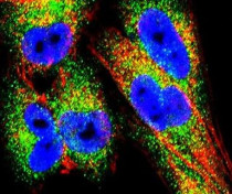
ARG55639 anti-TIMP1 antibody ICC/IF image
Immunofluorescence: A2058 cells stained with ARG55639 anti-TIMP1 antibody (green). Actin filaments have been labeled with Alexa Fluor 555 phalloidin (red). DAPI (blue) for nuclear staining.
-
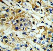
ARG55639 anti-TIMP1 antibody IHC-P image
Immunohistochemistry: Formalin-fixed and paraffin-embedded Human breast carcinoma stained with ARG55639 anti-TIMP1 antibody.
-
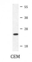
ARG55639 anti-TIMP1 antibody WB image
Western blot: 35 µg of CEM cell lysate stained with ARG55639 anti-TIMP1 antibody.
-
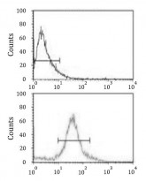
ARG55639 anti-TIMP1 antibody FACS image
Flow Cytometry: MDA-231 cells stained with ARG55639 anti-TIMP1 antibody (bottom histogram) or without primary antibody as control (top histogram), followed by incubation with FITC labelled secondary antibody.


