ARG63927
anti-TIM3 antibody
anti-TIM3 antibody for Flow cytometry,ICC/IF,Western blot and Human
Immune System antibody
Overview
| Product Description | Goat Polyclonal antibody recognizes TIM3 |
|---|---|
| Tested Reactivity | Hu |
| Predict Reactivity | Dog |
| Tested Application | FACS, ICC/IF, WB |
| Host | Goat |
| Clonality | Polyclonal |
| Isotype | IgG |
| Target Name | TIM3 |
| Antigen Species | Human |
| Immunogen | C-KWYSHSKEKIQN |
| Conjugation | Un-conjugated |
| Alternate Names | KIM-3; TIM3; TIMD-3; Tim-3; TIM-3; CD366; T-cell immunoglobulin and mucin domain-containing protein 3; TIMD3; T-cell membrane protein 3; HAVcr-2; T-cell immunoglobulin mucin receptor 3; Hepatitis A virus cellular receptor 2 |
Application Instructions
| Application Suggestion |
|
||||||||
|---|---|---|---|---|---|---|---|---|---|
| Application Note | WB: Recommend incubate at RT for 1h. * The dilutions indicate recommended starting dilutions and the optimal dilutions or concentrations should be determined by the scientist. |
Properties
| Form | Liquid |
|---|---|
| Purification | Purified from goat serum by antigen affinity chromatography. |
| Buffer | Tris saline (pH 7.3), 0.02% Sodium azide and 0.5% BSA. |
| Preservative | 0.02% Sodium azide |
| Stabilizer | 0.5% BSA |
| Concentration | 0.5 mg/ml |
| Storage Instruction | For continuous use, store undiluted antibody at 2-8°C for up to a week. For long-term storage, aliquot and store at -20°C or below. Storage in frost free freezers is not recommended. Avoid repeated freeze/thaw cycles. Suggest spin the vial prior to opening. The antibody solution should be gently mixed before use. |
| Note | For laboratory research only, not for drug, diagnostic or other use. |
Bioinformation
| Database Links |
Swiss-port # Q8TDQ0 Human Hepatitis A virus cellular receptor 2 |
|---|---|
| Gene Symbol | HAVCR2 |
| Background | The protein encoded by this gene belongs to the immunoglobulin superfamily, and TIM family of proteins. CD4-positive T helper lymphocytes can be divided into types 1 (Th1) and 2 (Th2) on the basis of their cytokine secretion patterns. Th1 cells are involved in cell-mediated immunity to intracellular pathogens and delayed-type hypersensitivity reactions, whereas, Th2 cells are involved in the control of extracellular helminthic infections and the promotion of atopic and allergic diseases. This protein is a Th1-specific cell surface protein that regulates macrophage activation, and inhibits Th1-mediated auto- and alloimmune responses, and promotes immunological tolerance. [provided by RefSeq, Sep 2011] |
| Research Area | Immune System antibody |
| Calculated MW | 33 kDa |
| PTM | O-glycosylated with core 1 or possibly core 8 glycans. Phosphorylated on tyrosine residues; modestly increased after TCR/CD28 stimulation. Can be phosphorylated in the cytoplasmatic domain by FYN (By similarity). Phosphorylation at Tyr-265 is increased by stimulation with ligand LGALS9. |
Images (5) Click the Picture to Zoom In
-
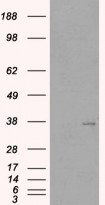
ARG63927 anti-TIM3 antibody WB image
Western blot: 1). Mock transfection; 2) HAVCR2 (RC209440) expressing plasmid transfected HEK293 cell lysate standed with ARG63927 anti-TIM3 antibody.
-
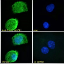
ARG63927 anti-TIM3 antibody ICC/IF image
Immunofluorescence: Paraformaldehyde fixed HepG2 cells permeabilized with 0.15% Triton. Cells were stained with ARG63927 anti-TIM3 antibody (green) at 10 µg/ml dilution for 1 hour. DAPI (blue) for nuclear staining. Negative control: Unimmunized goat IgG (green) at 10 µg/ml dilution.
-
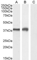
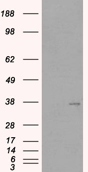
ARG63927 anti-TIM3 antibody WB image
Western blot: 35 µg of Jurkat (A), MOLT-4 (B) and A431 (C, negative control) cell lysates (in RIPA buffer) stained with ARG63927 anti-TIM3 antibody at 2 µg/ml dilution and incubated at RT for 1 hour.
-
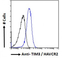
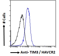
ARG63927 anti-TIM3 antibody FACS image
Flow Cytometry: Paraformaldehyde-fixed HepG2 cells permeabilized with 0.5% Triton. Cells were stained with ARG63927 anti-TIM3 antibody (blue line) at 10 µg/ml dilution for 1 hour, followed by incubation with Alexa FluorR 488 labelled secondary antibody. IgG control: Unimmunized goat IgG (black line).
-
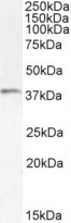
ARG63927 anti-TIM3 antibody WB image
Western blot: 35 µg of Human tonsil lysate (in RIPA buffer) stained with ARG63927 anti-TIM3 antibody at 1 µg/ml dilution and incubated at RT for 1 hour.