ARG56958
anti-TDP43 antibody [k1B8]
anti-TDP43 antibody [k1B8] for Flow cytometry,IHC-Formalin-fixed paraffin-embedded sections,Western blot and Human
Overview
| Product Description | Mouse Monoclonal antibody [k1B8] recognizes TDP43 |
|---|---|
| Tested Reactivity | Hu |
| Tested Application | FACS, IHC-P, WB |
| Host | Mouse |
| Clonality | Monoclonal |
| Clone | k1B8 |
| Isotype | IgG1, kappa |
| Target Name | TDP43 |
| Antigen Species | Human |
| Immunogen | Recombinant fragment around aa. 1-260 of Human TDP43. |
| Conjugation | Un-conjugated |
| Alternate Names | TAR DNA-binding protein 43; TDP-43; ALS10 |
Application Instructions
| Application Suggestion |
|
||||||||
|---|---|---|---|---|---|---|---|---|---|
| Application Note | IHC-P: Antigen Retrieval: Boil tissue section in 0.1M Sodium citrate buffer (pH 6.0) for 20 min. * The dilutions indicate recommended starting dilutions and the optimal dilutions or concentrations should be determined by the scientist. |
Properties
| Form | Liquid |
|---|---|
| Purification | Purification with Protein G. |
| Buffer | PBS (pH 7.4), 0.02% Sodium azide and 10% Glycerol. |
| Preservative | 0.02% Sodium azide |
| Stabilizer | 10% Glycerol |
| Concentration | 1 mg/ml |
| Storage Instruction | For continuous use, store undiluted antibody at 2-8°C for up to a week. For long-term storage, aliquot and store at -20°C. Storage in frost free freezers is not recommended. Avoid repeated freeze/thaw cycles. Suggest spin the vial prior to opening. The antibody solution should be gently mixed before use. |
| Note | For laboratory research only, not for drug, diagnostic or other use. |
Bioinformation
| Database Links | |
|---|---|
| Gene Symbol | TARDBP |
| Gene Full Name | TAR DNA binding protein |
| Background | HIV-1, the causative agent of acquired immunodeficiency syndrome (AIDS), contains an RNA genome that produces a chromosomally integrated DNA during the replicative cycle. Activation of HIV-1 gene expression by the transactivator Tat is dependent on an RNA regulatory element (TAR) located downstream of the transcription initiation site. The protein encoded by this gene is a transcriptional repressor that binds to chromosomally integrated TAR DNA and represses HIV-1 transcription. In addition, this protein regulates alternate splicing of the CFTR gene. A similar pseudogene is present on chromosome 20. [provided by RefSeq, Jul 2008] |
| Function | DNA and RNA-binding protein which regulates transcription and splicing. Involved in the regulation of CFTR splicing. It promotes CFTR exon 9 skipping by binding to the UG repeated motifs in the polymorphic region near the 3'-splice site of this exon. The resulting aberrant splicing is associated with pathological features typical of cystic fibrosis. May also be involved in microRNA biogenesis, apoptosis and cell division. Can repress HIV-1 transcription by binding to the HIV-1 long terminal repeat. Stabilizes the low molecular weight neurofilament (NFL) mRNA through a direct interaction with the 3' UTR. [UniProt] |
| Calculated MW | 45 kDa |
| PTM | Hyperphosphorylated in hippocampus, neocortex, and spinal cord from individuals affected with ALS and FTLDU. Ubiquitinated in hippocampus, neocortex, and spinal cord from individuals affected with ALS and FTLDU. Cleaved to generate C-terminal fragments in hippocampus, neocortex, and spinal cord from individuals affected with ALS and FTLDU. |
Images (4) Click the Picture to Zoom In
-

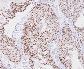
ARG56958 anti-TDP43 antibody [k1B8] IHC-P image
Immunohistochemistry: Paraffin embedded sections of Human prostate cancer tissue stained with ARG56958 anti-TDP43 antibody [k1B8] at 1:50 for 2 hours at RT. Antigen Retrieval: Boil tissue section in 0.1M Sodium citrate buffer (pH 6.0) for 20 min.
-

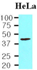
ARG56958 anti-TDP43 antibody [k1B8] WB image
Western blot: 20 µg of HeLa cell lysate stained with ARG56958 anti-TDP43 antibody [k1B8] at 1:1000.
-
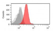
ARG56958 anti-TDP43 antibody [k1B8] FACS image
Flow Cytometry: HeLa cells stained with ARG56958 anti-TDP43 antibody [k1B8] at 2 - 5 µg/10^6 cells (red), followed by incubation with Alexa Fluor® 488 labelled secondary antibody. Mouse monoclonal IgG was used as the isotype control (dark gray), cells without incubation with primary and secondary antibody was used as the negative control (light gray).
-
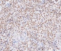
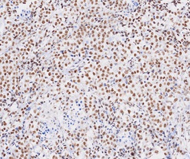
ARG56958 anti-TDP43 antibody [k1B8] IHC-P image
Immunohistochemistry: Paraffin embedded sections of Human seminoma tissue stained with ARG56958 anti-TDP43 antibody [k1B8] at 1:50 for 2 hours at RT. Antigen Retrieval: Boil tissue section in 0.1M Sodium citrate buffer (pH 6.0) for 20 min.