ARG10763
anti-TDP43 antibody [3H8]
anti-TDP43 antibody [3H8] for ICC/IF,IHC-Frozen sections,IHC-Formalin-fixed paraffin-embedded sections,Western blot and Human,Mouse,Rat
Overview
| Product Description | Mouse Monoclonal to Anti-Tar DNA Binding Protein 43 (TARBDP) |
|---|---|
| Tested Reactivity | Hu, Ms, Rat |
| Tested Application | ICC/IF, IHC-Fr, IHC-P, WB |
| Host | Mouse |
| Clonality | Monoclonal |
| Clone | 3H8 |
| Isotype | IgG1 |
| Target Name | TDP43 |
| Antigen Species | Human |
| Immunogen | Full length recombinant Human TDP43 expressed in and purified from E. coli. |
| Conjugation | Un-conjugated |
| Alternate Names | TAR DNA-binding protein 43; TDP-43; ALS10 |
Application Instructions
| Application Suggestion |
|
||||||||||
|---|---|---|---|---|---|---|---|---|---|---|---|
| Application Note | * The dilutions indicate recommended starting dilutions and the optimal dilutions or concentrations should be determined by the scientist. |
Properties
| Form | Liquid |
|---|---|
| Purification | Affinity purification. |
| Buffer | PBS and 50% Glycerol. |
| Stabilizer | 50% Glycerol |
| Concentration | 1 mg/ml |
| Storage Instruction | For continuous use, store undiluted antibody at 2-8°C for up to a week. For long-term storage, aliquot and store at -20°C. Storage in frost free freezers is not recommended. Avoid repeated freeze/thaw cycles. Suggest spin the vial prior to opening. The antibody solution should be gently mixed before use. |
| Note | For laboratory research only, not for drug, diagnostic or other use. |
Bioinformation
| Database Links | |
|---|---|
| Gene Symbol | TARDBP |
| Gene Full Name | TAR DNA binding protein |
| Background | HIV-1, the causative agent of acquired immunodeficiency syndrome (AIDS), contains an RNA genome that produces a chromosomally integrated DNA during the replicative cycle. Activation of HIV-1 gene expression by the transactivator Tat is dependent on an RNA regulatory element (TAR) located downstream of the transcription initiation site. The protein encoded by this gene is a transcriptional repressor that binds to chromosomally integrated TAR DNA and represses HIV-1 transcription. In addition, this protein regulates alternate splicing of the CFTR gene. A similar pseudogene is present on chromosome 20. [provided by RefSeq, Jul 2008] |
| Function | DNA and RNA-binding protein which regulates transcription and splicing. Involved in the regulation of CFTR splicing. It promotes CFTR exon 9 skipping by binding to the UG repeated motifs in the polymorphic region near the 3'-splice site of this exon. The resulting aberrant splicing is associated with pathological features typical of cystic fibrosis. May also be involved in microRNA biogenesis, apoptosis and cell division. Can repress HIV-1 transcription by binding to the HIV-1 long terminal repeat. Stabilizes the low molecular weight neurofilament (NFL) mRNA through a direct interaction with the 3' UTR. [UniProt] |
| Calculated MW | 45 kDa |
| PTM | Hyperphosphorylated in hippocampus, neocortex, and spinal cord from individuals affected with ALS and FTLDU. Ubiquitinated in hippocampus, neocortex, and spinal cord from individuals affected with ALS and FTLDU. Cleaved to generate C-terminal fragments in hippocampus, neocortex, and spinal cord from individuals affected with ALS and FTLDU. |
Images (4) Click the Picture to Zoom In
-
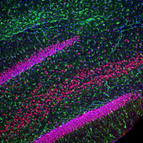
ARG10763 anti-TDP43 antibody [3H8] IHC-Fr image
Immunohistochemistry: Frozen section of Rat hippocampus stained with ARG10763 anti-TDP43 antibody [3H8] (red) at 1:2000 dilution and costained with ARG52313 anti-GFAP antibody (green) at 1:5000 dilution. DAPI (blue) for nuclear staining. (Sample preparation: Following transcardial perfusion of Rat with 4% paraformaldehyde, brain was post fixed for 24 hours, cut to 45 µM, and free-floating sections were stained with above antibodies.)
The TDP43 protein is concentrated in the nuclei of hippocampal neurons, while the GFAP antibody stains the intermediate filament network of astroglial cells.
-
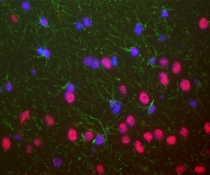
ARG10763 anti-TDP43 antibody [3H8] IHC-Fr image
Immunohistochemistry: Frozen sections of Formalin-fixed adult Rat brain, specifically the hippocampus. Hippocampal neuron nuclei are stained with ARG10763 anti-TDP43 antibody [3H8]. Chicken antibody to GFAP (green) shows the processes of astrocytic glial cells. Nuclei of all cells are revealed with DAPI DNA stain (blue). The TARDP antibody stains neuronal nuclei strongly and the nuclei of some non-neuronal cells much more weakly. Neuronal nuclei therefore look crimson, since they are both red due to the content of TDP43 and blue due to their content of DNA, stained blue with DAPI.
-
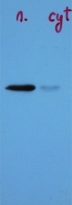
ARG10763 anti-TDP43 antibody [3H8] WB image
Western blot: Crude extract of Mouse brain nuclear fraction (left) and cytoplasmic fraction (right) stained with ARG10763 anti-TDP43 antibody [3H8]. There is a strong clear band in the nuclear preparation running at 43 kDa.
-
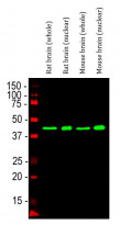
ARG10763 anti-TDP43 antibody [3H8] WB image
Western blot: Rat brain (whole), Rat brain (nuclear), Mouse brain (whole), Mouse brain (nuclear). The blots were stained with ARG10763 anti-TDP43 antibody [3H8] (green) at 1:2000 dilution.


