ARG66191
anti-TAK1 / MAP3K7 antibody
anti-TAK1 / MAP3K7 antibody for IHC-Formalin-fixed paraffin-embedded sections,Western blot and Human,Mouse,Rat
Overview
| Product Description | Rabbit Polyclonal antibody recognizes TAK1 / MAP3K7 |
|---|---|
| Tested Reactivity | Hu, Ms, Rat |
| Tested Application | IHC-P, WB |
| Specificity | The antibody detects endogenous Tak1 protein. |
| Host | Rabbit |
| Clonality | Polyclonal |
| Isotype | IgG |
| Target Name | TAK1 / MAP3K7 |
| Antigen Species | Human |
| Immunogen | Synthetic peptide around aa. 130-210 of Human TAK1 / MAP3K7. |
| Conjugation | Un-conjugated |
| Alternate Names | TAK1; Mitogen-activated protein kinase kinase kinase 7; EC 2.7.11.25; Transforming growth factor-beta-activated kinase 1; TGF-beta-activated kinase 1; MEKK7; TGF1a |
Application Instructions
| Application Suggestion |
|
||||||
|---|---|---|---|---|---|---|---|
| Application Note | IHC-P: Antigen Retrieval: Boil tissue section in Sodium citrate buffer (pH 6.0) for 20 min. * The dilutions indicate recommended starting dilutions and the optimal dilutions or concentrations should be determined by the scientist. |
Properties
| Form | Liquid |
|---|---|
| Purification | Affinity purification with immunogen. |
| Buffer | PBS, 0.02% Sodium azide, 50% Glycerol and 0.5% BSA. |
| Preservative | 0.02% Sodium azide |
| Stabilizer | 50% Glycerol and 0.5% BSA |
| Concentration | 1 mg/ml |
| Storage Instruction | For continuous use, store undiluted antibody at 2-8°C for up to a week. For long-term storage, aliquot and store at -20°C. Storage in frost free freezers is not recommended. Avoid repeated freeze/thaw cycles. Suggest spin the vial prior to opening. The antibody solution should be gently mixed before use. |
| Note | For laboratory research only, not for drug, diagnostic or other use. |
Bioinformation
| Database Links | |
|---|---|
| Gene Symbol | MAP3K7 |
| Gene Full Name | mitogen-activated protein kinase kinase kinase 7 |
| Background | The protein encoded by this gene is a member of the serine/threonine protein kinase family. This kinase mediates the signaling transduction induced by TGF beta and morphogenetic protein (BMP), and controls a variety of cell functions including transcription regulation and apoptosis. In response to IL-1, this protein forms a kinase complex including TRAF6, MAP3K7P1/TAB1 and MAP3K7P2/TAB2; this complex is required for the activation of nuclear factor kappa B. This kinase can also activate MAPK8/JNK, MAP2K4/MKK4, and thus plays a role in the cell response to environmental stresses. Four alternatively spliced transcript variants encoding distinct isoforms have been reported. [provided by RefSeq, Jul 2008] |
| Function | Serine/threonine kinase which acts as an essential component of the MAP kinase signal transduction pathway. Plays an important role in the cascades of cellular responses evoked by changes in the environment. Mediates signal transduction of TRAF6, various cytokines including interleukin-1 (IL-1), transforming growth factor-beta (TGFB), TGFB-related factors like BMP2 and BMP4, toll-like receptors (TLR), tumor necrosis factor receptor CD40 and B-cell receptor (BCR). Ceramides are also able to activate MAP3K7/TAK1. Once activated, acts as an upstream activator of the MKK/JNK signal transduction cascade and the p38 MAPK signal transduction cascade through the phosphorylation and activation of several MAP kinase kinases like MAP2K1/MEK1, MAP2K3/MKK3, MAP2K6/MKK6 and MAP2K7/MKK7. These MAP2Ks in turn activate p38 MAPKs, c-jun N-terminal kinases (JNKs) and I-kappa-B kinase complex (IKK). Both p38 MAPK and JNK pathways control the transcription factors activator protein-1 (AP-1), while nuclear factor-kappa B is activated by IKK. MAP3K7 activates also IKBKB and MAPK8/JNK1 in response to TRAF6 signaling and mediates BMP2-induced apoptosis. In osmotic stress signaling, plays a major role in the activation of MAPK8/JNK1, but not that of NF-kappa-B. Promotes TRIM5 capsid-specific restriction activity. [UniProt] |
| Calculated MW | 67 kDa |
| PTM | Association with TAB1/MAP3K7IP1 promotes autophosphorylation at Ser-192 and subsequent activation. Association with TAB2/MAP3K7IP2, itself associated with free unanchored Lys-63 polyubiquitin chain, promotes autophosphorylation and subsequent activation of MAP3K7. Dephosphorylation at Ser-192 by PPM1B/PP2CB and at Thr-187 by PP2A and PPP6C leads to inactivation. 'Lys-48'-linked polyubiquitination at Lys-72 is induced by TNFalpha, and leads to proteasomal degradation. Undergoes 'Lys-48'-linked polyubiquitination catalyzed by ITCH (By similarity). Requires 'Lys-63'-linked polyubiquitination for autophosphorylation and subsequent activation. 'Lys-63'-linked ubiquitination does not lead to proteasomal degradation. Deubiquitinated by CYLD, a protease that selectively cleaves 'Lys-63'-linked ubiquitin chains. Deubiquitinated by Y.enterocolitica YopP. |
Images (12) Click the Picture to Zoom In
-
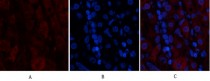
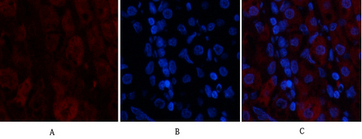
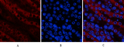
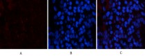
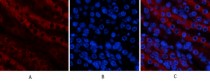
ARG66191 anti-TAK1 / MAP3K7 antibody IHC image
Immunohistochemistry: Human stomach tissue stained with ARG66191 anti-TAK1 / MAP3K7 antibody (red) at 1:200 dilution (4°C, overnight).
Picture A: Target. Picture B: DAPI. Picture C: merge of A+B.
-
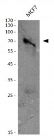
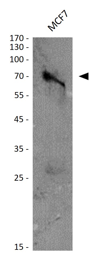
ARG66191 anti-TAK1 / MAP3K7 antibody WB image
Western blot: MCF7 cell lysate stained with ARG66191 anti-TAK1 / MAP3K7 antibody at 1:1000 dilution.
-
ARG66191 anti-TAK1 / MAP3K7 antibody IHC image
Immunohistochemistry: Human stomach tissue stained with ARG66191 anti-TAK1 / MAP3K7 antibody (red) at 1:200 dilution (4°C, overnight).
Picture A: Target. Picture B: DAPI. Picture C: merge of A+B.
-
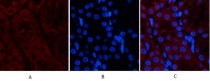
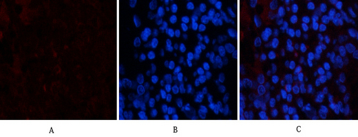
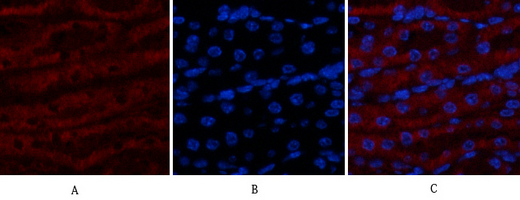
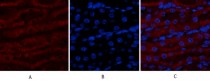
ARG66191 anti-TAK1 / MAP3K7 antibody IHC image
Immunohistochemistry: Rat kidney tissue stained with ARG66191 anti-TAK1 / MAP3K7 antibody (red) at 1:200 dilution (4°C, overnight).
Picture A: Target. Picture B: DAPI. Picture C: merge of A+B.
-
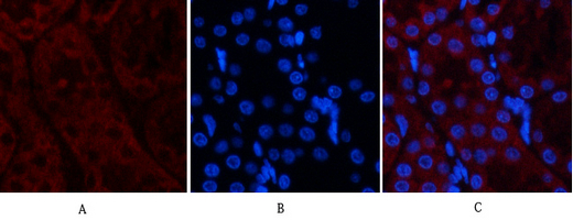
ARG66191 anti-TAK1 / MAP3K7 antibody IHC image
Immunohistochemistry: Rat kidney tissue stained with ARG66191 anti-TAK1 / MAP3K7 antibody (red) at 1:200 dilution (4°C, overnight).
Picture A: Target. Picture B: DAPI. Picture C: merge of A+B.
-
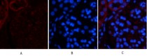
ARG66191 anti-TAK1 / MAP3K7 antibody IHC image
Immunohistochemistry: Mouse kidney tissue stained with ARG66191 anti-TAK1 / MAP3K7 antibody (red) at 1:200 dilution (4°C, overnight).
Picture A: Target. Picture B: DAPI. Picture C: merge of A+B.
-
ARG66191 anti-TAK1 / MAP3K7 antibody IHC image
Immunohistochemistry: Mouse kidney tissue stained with ARG66191 anti-TAK1 / MAP3K7 antibody (red) at 1:200 dilution (4°C, overnight).
Picture A: Target. Picture B: DAPI. Picture C: merge of A+B.
-
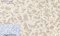
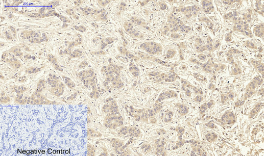
ARG66191 anti-TAK1 / MAP3K7 antibody IHC-P image
Immunohistochemistry: Paraffin-embedded Human liver cancer tissue stained with ARG66191 anti-TAK1 / MAP3K7 antibody at 1:200 dilution (4°C, overnight). Antigen Retrieval: Boil tissue section in Sodium citrate buffer (pH 6.0) for 20 min.
Negative control was used by secondary antibody only.
-

ARG66191 anti-TAK1 / MAP3K7 antibody IHC-P image
Immunohistochemistry: Paraffin-embedded Human stomach tissue stained with ARG66191 anti-TAK1 / MAP3K7 antibody at 1:200 dilution (4°C, overnight). Antigen Retrieval: Boil tissue section in Sodium citrate buffer (pH 6.0) for 20 min.
Negative control was used by secondary antibody only.
-
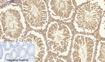
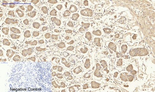
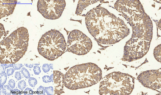
ARG66191 anti-TAK1 / MAP3K7 antibody IHC-P image
Immunohistochemistry: Paraffin-embedded Rat testis tissue stained with ARG66191 anti-TAK1 / MAP3K7 antibody at 1:200 dilution (4°C, overnight). Antigen Retrieval: Boil tissue section in Sodium citrate buffer (pH 6.0) for 20 min.
Negative control was used by secondary antibody only.
-
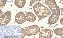
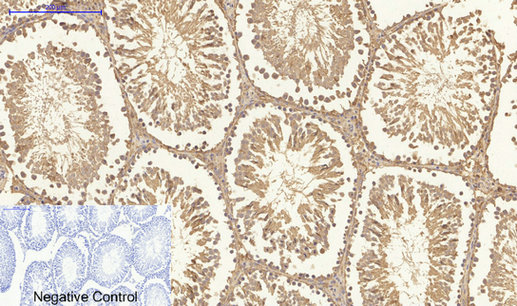
ARG66191 anti-TAK1 / MAP3K7 antibody IHC-P image
Immunohistochemistry: Paraffin-embedded Mouse testis tissue stained with ARG66191 anti-TAK1 / MAP3K7 antibody at 1:200 dilution (4°C, overnight). Antigen Retrieval: Boil tissue section in Sodium citrate buffer (pH 6.0) for 20 min.
Negative control was used by secondary antibody only.
-
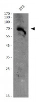
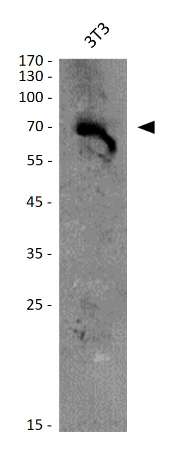
ARG66191 anti-TAK1 / MAP3K7 antibody WB image
Western blot: 3T3 cell lysate stained with ARG66191 anti-TAK1 / MAP3K7 antibody at 1:1000 dilution.