ARG58134
anti-Stathmin 1 antibody
anti-Stathmin 1 antibody for Flow cytometry,ICC/IF,IHC-Formalin-fixed paraffin-embedded sections,Western blot and Human,Mouse,Rat
Overview
| Product Description | Rabbit Polyclonal antibody recognizes Stathmin 1 |
|---|---|
| Tested Reactivity | Hu, Ms, Rat |
| Tested Application | FACS, ICC/IF, IHC-P, WB |
| Host | Rabbit |
| Clonality | Polyclonal |
| Isotype | IgG |
| Target Name | Stathmin 1 |
| Antigen Species | Human |
| Immunogen | Synthetic peptide of Human Stathmin 1. (ASSDIQVKELEKRASGQAFELILSPRSKESVPE) |
| Conjugation | Un-conjugated |
| Alternate Names | PP17; Prosolin; Stathmin; Protein Pr22; PR22; Lag; C1orf215; PP19; pp19; SMN; OP18; Leukemia-associated phosphoprotein p18; LAP18; pp17; Oncoprotein 18; Phosphoprotein p19; Op18; Metablastin |
Application Instructions
| Application Suggestion |
|
||||||||||
|---|---|---|---|---|---|---|---|---|---|---|---|
| Application Note | IHC-P: Antigen Retrieval: Boil tissue section in 10 mM Citrate buffer (pH 6.0) for 20 min or EDTA buffer (pH 8.0) for 20 min, followed by cooling at RT. * The dilutions indicate recommended starting dilutions and the optimal dilutions or concentrations should be determined by the scientist. |
||||||||||
| Positive Control | Rat brain, Rat testis, Mouse brain, Mouse testis, MDA-MB-453, SH-SY5Y and Raji | ||||||||||
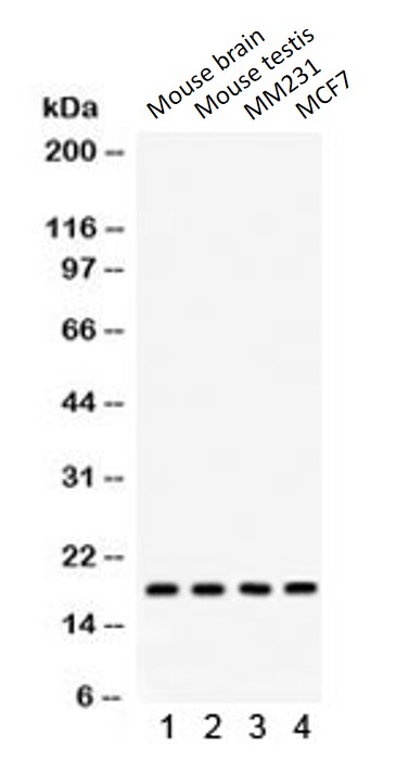
| Observed Size | ~ 20 kDa |
Properties
| Form | Liquid |
|---|---|
| Purification | Affinity purification with immunogen. |
| Buffer | PBS, 0.025% Sodium azide and 2.5% BSA. |
| Preservative | 0.025% Sodium azide |
| Stabilizer | 2.5% BSA |
| Concentration | 0.5 mg/ml |
| Storage Instruction | For continuous use, store undiluted antibody at 2-8°C for up to a week. For long-term storage, aliquot and store at -20°C or below. Storage in frost free freezers is not recommended. Avoid repeated freeze/thaw cycles. Suggest spin the vial prior to opening. The antibody solution should be gently mixed before use. |
| Note | For laboratory research only, not for drug, diagnostic or other use. |
Bioinformation
| Database Links | |
|---|---|
| Gene Symbol | STMN1 |
| Gene Full Name | stathmin 1 |
| Background | This gene belongs to the stathmin family of genes. It encodes a ubiquitous cytosolic phosphoprotein proposed to function as an intracellular relay integrating regulatory signals of the cellular environment. The encoded protein is involved in the regulation of the microtubule filament system by destabilizing microtubules. It prevents assembly and promotes disassembly of microtubules. Multiple transcript variants encoding different isoforms have been found for this gene. [provided by RefSeq, Feb 2009] |
| Function | Involved in the regulation of the microtubule (MT) filament system by destabilizing microtubules. Prevents assembly and promotes disassembly of microtubules. Phosphorylation at Ser-16 may be required for axon formation during neurogenesis. Involved in the control of the learned and innate fear (By similarity). [UniProt] |
| Cellular Localization | Cytoplasmic. [UniProt] |
| Calculated MW | 17 kDa |
| PTM | Many different phosphorylated forms are observed depending on specific combinations among the sites which can be phosphorylated. MAPK is responsible for the phosphorylation of stathmin in response to NGF. Phosphorylation at Ser-16 seems to be required for neuron polarization (By similarity). Phosphorylation at Ser-63 reduces tubulin binding 10-fold and suppresses the MT polymerization inhibition activity. [UniProt] |
Images (13) Click the Picture to Zoom In
-
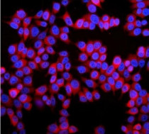
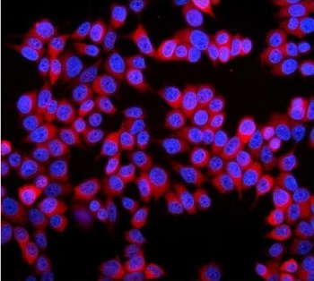
ARG58134 anti-Stathmin 1 antibody ICC/IF image
Immunofluorescence: MCF7 cells stained with ARG58134 anti-Stathmin 1 antibody (red). DAPI (blue) for nuclear staining.
-
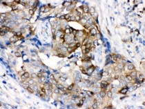
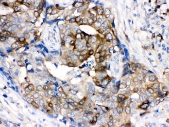
ARG58134 anti-Stathmin 1 antibody IHC-P image
Immunohistochemistry: Formalin-fixed and paraffin-embedded Human breast cancer tissue stained with ARG58134 anti-Stathmin 1 antibody. Antigen Retrieval: Boil tissue section in 10mM Citrate buffer (pH 6.0) for 20 min followed by cooling at RT.
-
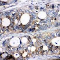
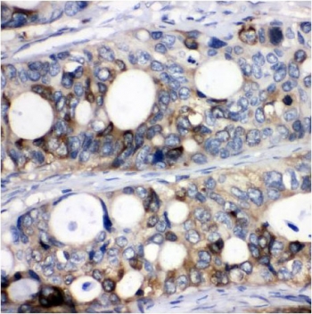
ARG58134 anti-Stathmin 1 antibody IHC-P image
Immunohistochemistry: Formalin-fixed and paraffin-embedded Mouse testis tissue. Antigen Retrieval: Boil tissue section in 10 mM Citrate buffer (pH 6.0) for 20 min, followed by cooling at RT. The tissue section was stained with ARG58134 anti-Stathmin 1 antibody.
-
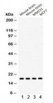
ARG58134 anti-Stathmin 1 antibody WB image
Western blot: Mouse brain, Mouse testis, MM231 and MCF7 cell lysates stained with ARG58134 anti-Stathmin 1 antibody.
-
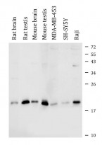
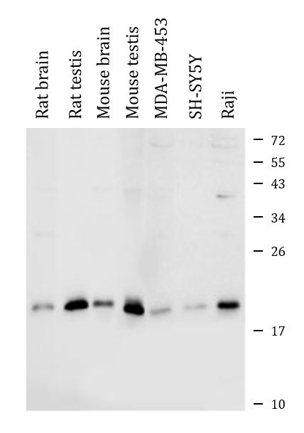
ARG58134 anti-Stathmin 1 antibody WB image
Western blot: Rat brain, Rat testis, Mouse brain, Mouse testis, MDA-MB-453, SH-SY5Y and Raji cell lysates stained with ARG58134 anti-Stathmin 1 antibody.
-
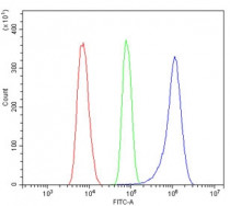
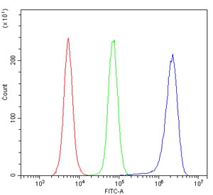
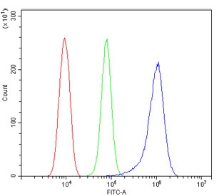
ARG58134 anti-Stathmin 1 antibody FACS image
Flow Cytometry: ThP-1 cells were blocked with goat sera and stained with ARG58134 anti-Stathmin 1 antibody at 1 µg/10^6 cells (blue); Cells alone (red); Isotype control (green).
-
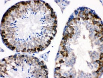
ARG58134 anti-Stathmin 1 antibody IHC-P image
Immunohistochemistry: Formalin-fixed and paraffin-embedded Mouse testis stained with ARG58134 anti-Stathmin 1 antibody. Antigen Retrieval: Boil tissue section in 10mM Citrate buffer (pH 6.0) for 20 min followed by cooling at RT.
-
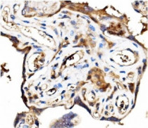
ARG58134 anti-Stathmin 1 antibody IHC-P image
Immunohistochemistry: Formalin-fixed and paraffin-embedded Human placental tissue. Antigen Retrieval: Boil tissue section in EDTA buffer (pH 8.0) for 20 min, followed by cooling at RT. The tissue section was stained with ARG58134 anti-Stathmin 1 antibody.
-
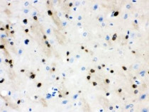
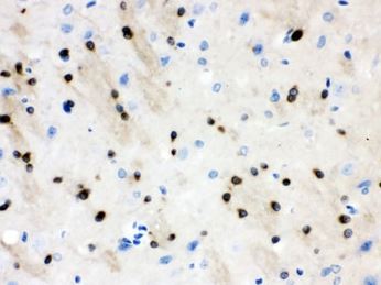
ARG58134 anti-Stathmin 1 antibody IHC-P image
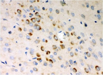
Immunohistochemistry: Formalin-fixed and paraffin-embedded Rat brain stained with ARG58134 anti-Stathmin 1 antibody. Antigen Retrieval: Boil tissue section in 10mM Citrate buffer (pH 6.0) for 20 min followed by cooling at RT.
-

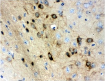
ARG58134 anti-Stathmin 1 antibody IHC-P image
Immunohistochemistry: Formalin-fixed and paraffin-embedded Mouse brain tissue. Antigen Retrieval: Boil tissue section in EDTA buffer (pH 8.0) for 20 min, followed by cooling at RT. The tissue section was stained with ARG58134 anti-Stathmin 1 antibody.
-
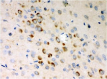
ARG58134 anti-Stathmin 1 antibody IHC-P image
Immunohistochemistry: Formalin-fixed and paraffin-embedded Rat brain tissue. Antigen Retrieval: Boil tissue section in EDTA buffer (pH 8.0) for 20 min, followed by cooling at RT. The tissue section was stained with ARG58134 anti-Stathmin 1 antibody.
-
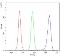
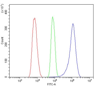
ARG58134 anti-Stathmin 1 antibody FACS image
Flow Cytometry: Raw264.7 cells were blocked with goat sera and stained with ARG58134 anti-Stathmin 1 antibody at 1 µg/10^6 cells (blue); Cells alone (red); Isotype control (green).
-
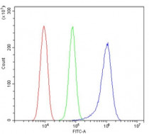
ARG58134 anti-Stathmin 1 antibody FACS image
Flow Cytometry: RH-35 cells were blocked with goat sera and stained with ARG58134 anti-Stathmin 1 antibody at 1 µg/10^6 cells (blue); Cells alone (red); Isotype control (green).