ARG51652
anti-Src phospho (Tyr529) antibody
anti-Src phospho (Tyr529) antibody for ICC/IF,IHC-Formalin-fixed paraffin-embedded sections,Western blot and Human,Mouse,Rat
Cancer antibody; Gene Regulation antibody; Signaling Transduction antibody; Src Family Protein Tyrosine Kinases antibody
Overview
| Product Description | Rabbit Polyclonal antibody recognizes Src phospho (Tyr529) |
|---|---|
| Tested Reactivity | Hu, Ms, Rat |
| Tested Application | ICC/IF, IHC-P, WB |
| Host | Rabbit |
| Clonality | Polyclonal |
| Isotype | IgG |
| Target Name | Src |
| Antigen Species | Human |
| Immunogen | Peptide sequence around phosphorylation site of tyrosine 529 (P-Q-Y(p)-Q-P) derived from Human Src. |
| Conjugation | Un-conjugated |
| Alternate Names | Proto-oncogene c-Src; ASV; p60-Src; c-SRC; Proto-oncogene tyrosine-protein kinase Src; SRC1; pp60c-src; EC 2.7.10.2 |
Application Instructions
| Application Suggestion |
|
||||||||
|---|---|---|---|---|---|---|---|---|---|
| Application Note | * The dilutions indicate recommended starting dilutions and the optimal dilutions or concentrations should be determined by the scientist. |
Properties
| Form | Liquid |
|---|---|
| Purification | Antibodies were produced by immunizing rabbits with KLH-conjugated synthetic phosphopeptide. Antibodies were purified by affinity-chromatography using epitope-specific phosphopeptide. In addition, non-phospho specific antibodies were removed by chromatogramphy using non-phosphopeptide. |
| Buffer | PBS (without Mg2+ and Ca2+, pH 7.4), 150mM NaCl, 0.02% Sodium azide and 50% Glycerol. |
| Preservative | 0.02% Sodium azide |
| Stabilizer | 50% Glycerol |
| Concentration | 1 mg/ml |
| Storage Instruction | For continuous use, store undiluted antibody at 2-8°C for up to a week. For long-term storage, aliquot and store at -20°C. Storage in frost free freezers is not recommended. Avoid repeated freeze/thaw cycles. Suggest spin the vial prior to opening. The antibody solution should be gently mixed before use. |
| Note | For laboratory research only, not for drug, diagnostic or other use. |
Bioinformation
| Database Links | |
|---|---|
| Gene Symbol | SRC |
| Gene Full Name | SRC proto-oncogene, non-receptor tyrosine kinase |
| Background | proto-oncogenic cytoplasmic tyrosine kinase of the SRC family. Highly expressed in certain fully differentiated cells such as neurons, platelets and macrophages. Phosphorylation of an activation loop tyrosine activates the enzyme; phosphorylation of a tyrosine in the C-terminus by Csk inhibits the enzyme. |
| Function | Non-receptor protein tyrosine kinase which is activated following engagement of many different classes of cellular receptors including immune response receptors, integrins and other adhesion receptors, receptor protein tyrosine kinases, G protein-coupled receptors as well as cytokine receptors. Participates in signaling pathways that control a diverse spectrum of biological activities including gene transcription, immune response, cell adhesion, cell cycle progression, apoptosis, migration, and transformation. Due to functional redundancy between members of the SRC kinase family, identification of the specific role of each SRC kinase is very difficult. SRC appears to be one of the primary kinases activated following engagement of receptors and plays a role in the activation of other protein tyrosine kinase (PTK) families. Receptor clustering or dimerization leads to recruitment of SRC to the receptor complexes where it phosphorylates the tyrosine residues within the receptor cytoplasmic domains. Plays an important role in the regulation of cytoskeletal organization through phosphorylation of specific substrates such as AFAP1. Phosphorylation of AFAP1 allows the SRC SH2 domain to bind AFAP1 and to localize to actin filaments. Cytoskeletal reorganization is also controlled through the phosphorylation of cortactin (CTTN). When cells adhere via focal adhesions to the extracellular matrix, signals are transmitted by integrins into the cell resulting in tyrosine phosphorylation of a number of focal adhesion proteins, including PTK2/FAK1 and paxillin (PXN). In addition to phosphorylating focal adhesion proteins, SRC is also active at the sites of cell-cell contact adherens junctions and phosphorylates substrates such as beta-catenin (CTNNB1), delta-catenin (CTNND1), and plakoglobin (JUP). Another type of cell-cell junction, the gap junction, is also a target for SRC, which phosphorylates connexin-43 (GJA1). SRC is implicated in regulation of pre-mRNA-processing and phosphorylates RNA-binding proteins such as KHDRBS1. Also plays a role in PDGF-mediated tyrosine phosphorylation of both STAT1 and STAT3, leading to increased DNA binding activity of these transcription factors. Involved in the RAS pathway through phosphorylation of RASA1 and RASGRF1. Plays a role in EGF-mediated calcium-activated chloride channel activation. Required for epidermal growth factor receptor (EGFR) internalization through phosphorylation of clathrin heavy chain (CLTC and CLTCL1) at 'Tyr-1477'. Involved in beta-arrestin (ARRB1 and ARRB2) desensitization through phosphorylation and activation of ADRBK1, leading to beta-arrestin phosphorylation and internalization. Has a critical role in the stimulation of the CDK20/MAPK3 mitogen-activated protein kinase cascade by epidermal growth factor. Might be involved not only in mediating the transduction of mitogenic signals at the level of the plasma membrane but also in controlling progression through the cell cycle via interaction with regulatory proteins in the nucleus. Plays an important role in osteoclastic bone resorption in conjunction with PTK2B/PYK2. Both the formation of a SRC-PTK2B/PYK2 complex and SRC kinase activity are necessary for this function. Recruited to activated integrins by PTK2B/PYK2, thereby phosphorylating CBL, which in turn induces the activation and recruitment of phosphatidylinositol 3-kinase to the cell membrane in a signaling pathway that is critical for osteoclast function. Promotes energy production in osteoclasts by activating mitochondrial cytochrome C oxidase. Phosphorylates DDR2 on tyrosine residues, thereby promoting its subsequent autophosphorylation. Phosphorylates RUNX3 and COX2 on tyrosine residues, TNK2 on 'Tyr-284' and CBL on 'Tyr-731'. Enhances DDX58/RIG-I-elicited antiviral signaling. Phosphorylates PDPK1 at 'Tyr-9', 'Tyr-373' and 'Tyr-376'. Phosphorylates BCAR1 at 'Tyr-128'. Phosphorylates CBLC at multiple tyrosine residues, phosphorylation at 'Tyr-341' activates CBLC E3 activity. [UniProt] |
| Highlight | Related Antibody Duos and Panels: ARG30170 Phospho Src Antibody Panel Related products: Src antibodies; Src ELISA Kits; Src Duos / Panels; Anti-Rabbit IgG secondary antibodies; |
| Research Area | Cancer antibody; Gene Regulation antibody; Signaling Transduction antibody; Src Family Protein Tyrosine Kinases antibody |
| Calculated MW | 60 kDa |
| PTM | Myristoylated at Gly-2, and this is essential for targeting to membranes. Dephosphorylated at Tyr-530 by PTPRJ (By similarity). Phosphorylated on Tyr-530 by c-Src kinase (CSK). The phosphorylated form is termed pp60c-src. Dephosphorylated by PTPRJ at Tyr-419. Normally maintained in an inactive conformation with the SH2 domain engaged with Tyr-530, the SH3 domain engaged with the SH2-kinase linker, and Tyr-419 dephosphorylated. Dephosphorylation of Tyr-530 as a result of protein tyrosine phosphatase (PTP) action disrupts the intramolecular interaction between the SH2 domain and Tyr-530, Tyr-419 can then become autophosphorylated, resulting in SRC activation. Phosphorylation of Tyr-530 by CSK allows this interaction to reform, resulting in SRC inactivation. CDK5-mediated phosphorylation at Ser-75 targets SRC to ubiquitin-dependent degradation and thus leads to cytoskeletal reorganization. Phosphorylated by PTK2/FAK1; this enhances kinase activity. Phosphorylated by PTK2B/PYK2; this enhances kinase activity. S-nitrosylation is important for activation of its kinase activity. Ubiquitinated in response to CDK5-mediated phosphorylation. Ubiquitination mediated by CBLC requires SRC autophosphorylation at Tyr-419 and may lead to lysosomal degradation. |
Images (4) Click the Picture to Zoom In
-

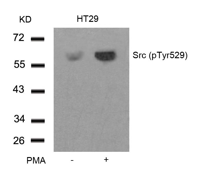
ARG51652 anti-Src phospho (Tyr529) antibody WB image
Western blot: Extracts from HT29 cells untreated or treated with PMA stained with ARG51652 anti-Src phospho (Tyr529) antibody.
-
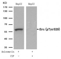
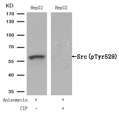
ARG51652 anti-Src phospho (Tyr529) antibody WB image
Western blot: Extracts from HepG2 cells, treated with Anisomycin or calf intestinal phosphatase (CIP), stained with ARG51652 anti-Src phospho (Tyr529) antibody.
-
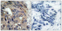
ARG51652 anti-Src phospho (Tyr529) antibody IHC-P image
Immunohistochemistry: Paraffin-embedded Human breast carcinoma tissue stained with ARG51652 anti-Src phospho (Tyr529) antibody (left) or the same antibody preincubated with blocking peptide (right).
-

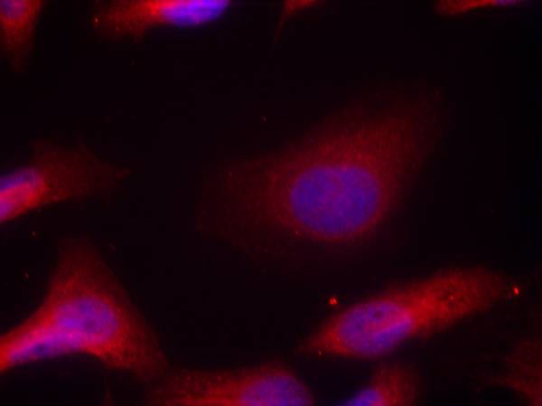
ARG51652 anti-Src phospho (Tyr529) antibody ICC/IF image
Immunofluorescence: methanol-fixed HeLa cells stained with ARG51652 anti-Src phospho (Tyr529) antibody.