ARG42203
anti-Smad 7 antibody
anti-Smad 7 antibody for ICC/IF,Western blot and Human,Mouse,Rat
Overview
| Product Description | Rabbit Polyclonal antibody recognizes Smad 7 |
|---|---|
| Tested Reactivity | Hu, Ms, Rat |
| Tested Application | ICC/IF, WB |
| Host | Rabbit |
| Clonality | Polyclonal |
| Isotype | IgG |
| Target Name | Smad 7 |
| Antigen Species | Human |
| Immunogen | Synthetic peptide within aa. 1-100 of Human Smad 7 (NP_005895.1). |
| Conjugation | Un-conjugated |
| Alternate Names | Mothers against decapentaplegic homolog 8; MADH8; MADH7; Smad7; Mothers against DPP homolog 7; Mothers against decapentaplegic homolog 7; hSMAD7; Mothers against DPP homolog 8; MAD homolog 8; CRCS3; SMAD 7; MAD homolog 7; SMAD family member 7 |
Application Instructions
| Application Suggestion |
|
||||||
|---|---|---|---|---|---|---|---|
| Application Note | * The dilutions indicate recommended starting dilutions and the optimal dilutions or concentrations should be determined by the scientist. | ||||||
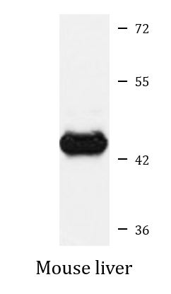
| Positive Control | Mouse liver | ||||||
| Observed Size | ~ 43 kDa |
Properties
| Form | Liquid |
|---|---|
| Purification | Affinity purified. |
| Buffer | PBS (pH 7.3), 0.02% Sodium azide and 50% Glycerol. |
| Preservative | 0.02% Sodium azide |
| Stabilizer | 50% Glycerol |
| Storage Instruction | For continuous use, store undiluted antibody at 2-8°C for up to a week. For long-term storage, aliquot and store at -20°C. Storage in frost free freezers is not recommended. Avoid repeated freeze/thaw cycles. Suggest spin the vial prior to opening. The antibody solution should be gently mixed before use. |
| Note | For laboratory research only, not for drug, diagnostic or other use. |
Bioinformation
| Database Links | |
|---|---|
| Gene Symbol | SMAD7 |
| Gene Full Name | SMAD family member 7 |
| Background | The protein encoded by this gene is a nuclear protein that binds the E3 ubiquitin ligase SMURF2. Upon binding, this complex translocates to the cytoplasm, where it interacts with TGF-beta receptor type-1 (TGFBR1), leading to the degradation of both the encoded protein and TGFBR1. Expression of this gene is induced by TGFBR1. Variations in this gene are a cause of susceptibility to colorectal cancer type 3 (CRCS3). Several transcript variants encoding different isoforms have been found for this gene. [provided by RefSeq, Jun 2010] |
| Function | Antagonist of signaling by TGF-beta (transforming growth factor) type 1 receptor superfamily members; has been shown to inhibit TGF-beta (Transforming growth factor) and activin signaling by associating with their receptors thus preventing SMAD2 access. Functions as an adapter to recruit SMURF2 to the TGF-beta receptor complex. Also acts by recruiting the PPP1R15A-PP1 complex to TGFBR1, which promotes its dephosphorylation. Positively regulates PDPK1 kinase activity by stimulating its dissociation from the 14-3-3 protein YWHAQ which acts as a negative regulator. [UniProt] |
| Cellular Localization | Nucleus. Cytoplasm. Note=Interaction with NEDD4L or RNF111 induces translocation from the nucleus to the cytoplasm (PubMed:16601693). TGF-beta stimulates its translocation from the nucleus to the cytoplasm. PDPK1 inhibits its translocation from the nucleus to the cytoplasm in response to TGF-beta (PubMed:17327236). [UniProt] |
| Calculated MW | 46 kDa |
| PTM | Phosphorylation on Ser-249 does not affect its stability, nuclear localization or inhibitory function in TGFB signaling; however it affects its ability to regulate transcription (By similarity). Phosphorylated by PDPK1. Ubiquitinated by WWP1 (By similarity). Polyubiquitinated by RNF111, which is enhanced by AXIN1 and promotes proteasomal degradation (PubMed:14657019, PubMed:16601693). In response to TGF-beta, ubiquitinated by SMURF1; which promotes its degradation (PubMed:11278251). Acetylation prevents ubiquitination and degradation mediated by SMURF1. [UniProt] |
Images (1) Click the Picture to Zoom In
-

ARG42203 anti-Smad 7 antibody WB image
Western blot: 25 µg of Mouse liver lysate stained with ARG42203 anti-Smad 7 antibody at 1:1000 dilution.
Specific References
Expression of SMADs in orthotopic human endometrium, ovarian endometriosis, and endometriotic lesions in a murine model
IHC-P / Mouse


