ARG43102
anti-Semaphorin 6A antibody
anti-Semaphorin 6A antibody for Flow cytometry,IHC-Formalin-fixed paraffin-embedded sections,Western blot and Human,Mouse,Rat
Overview
| Product Description | Rabbit Polyclonal antibody recognizes Semaphorin 6A |
|---|---|
| Tested Reactivity | Hu, Ms, Rat |
| Tested Application | FACS, IHC-P, WB |
| Host | Rabbit |
| Clonality | Polyclonal |
| Isotype | IgG |
| Target Name | Semaphorin 6A |
| Antigen Species | Human |
| Immunogen | Recombinant protein corresponding to H31-N306 of Human Semaphorin 6A. |
| Conjugation | Un-conjugated |
| Alternate Names | VIA; Semaphorin VIA; Semaphorin-6A; SEMAQ; Semaphorin-6A-1; SEMA; Sema VIA; SEMA6A-1; HT018; SEMA6A1 |
Application Instructions
| Application Suggestion |
|
||||||||
|---|---|---|---|---|---|---|---|---|---|
| Application Note | IHC-P: Antigen Retrieval: Heat mediation was performed in EDTA buffer (pH 8.0). * The dilutions indicate recommended starting dilutions and the optimal dilutions or concentrations should be determined by the scientist. |
||||||||
| Observed Size | ~ 110 kDa |
Properties
| Form | Liquid |
|---|---|
| Purification | Affinity purification with immunogen. |
| Buffer | 0.2% Na2HPO4, 0.9% NaCl, 0.01% Sodium azide and 4% Trehalose. |
| Preservative | 0.01% Sodium azide |
| Stabilizer | 4% Trehalose |
| Concentration | 0.5 mg/ml |
| Storage Instruction | For continuous use, store undiluted antibody at 2-8°C for up to a week. For long-term storage, aliquot and store at -20°C or below. Storage in frost free freezers is not recommended. Avoid repeated freeze/thaw cycles. Suggest spin the vial prior to opening. The antibody solution should be gently mixed before use. |
| Note | For laboratory research only, not for drug, diagnostic or other use. |
Bioinformation
| Database Links | |
|---|---|
| Gene Symbol | SEMA6A |
| Gene Full Name | sema domain, transmembrane domain (TM), and cytoplasmic domain, (semaphorin) 6A |
| Background | The transmembrane semaphorin SEMA6A is expressed in developing neural tissue and is required for proper development of the thalamocortical projection (Leighton et al., 2001 [PubMed 11242070]).[supplied by OMIM, Feb 2011] |
| Function | Cell surface receptor for PLXNA2 that plays an important role in cell-cell signaling. Required for normal granule cell migration in the developing cerebellum. Promotes reorganization of the actin cytoskeleton and plays an important role in axon guidance in the developing central nervous system. Can act as repulsive axon guidance cue. Has repulsive action towards migrating granular neurons. May play a role in channeling sympathetic axons into the sympathetic chains and controlling the temporal sequence of sympathetic target innervation (By similarity). [UniProt] |
| Cellular Localization | Cell membrane; Single-pass type I membrane protein. [UniProt] |
| Calculated MW | 114 kDa |
Images (3) Click the Picture to Zoom In
-
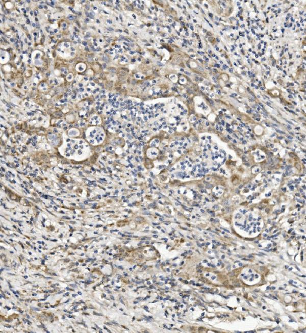
ARG43102 anti-Semaphorin 6A antibody IHC-P image
Immunohistochemistry: Paraffin-embedded Human rectal cancer tissue. Antigen Retrieval: Heat mediation was performed in EDTA buffer (pH 8.0). The tissue section was blocked with 10% goat serum. The tissue section was then stained with ARG43102 anti-Semaphorin 6A antibody at 1 µg/ml dilution, overnight at 4°C.
-
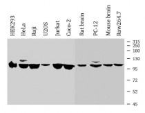
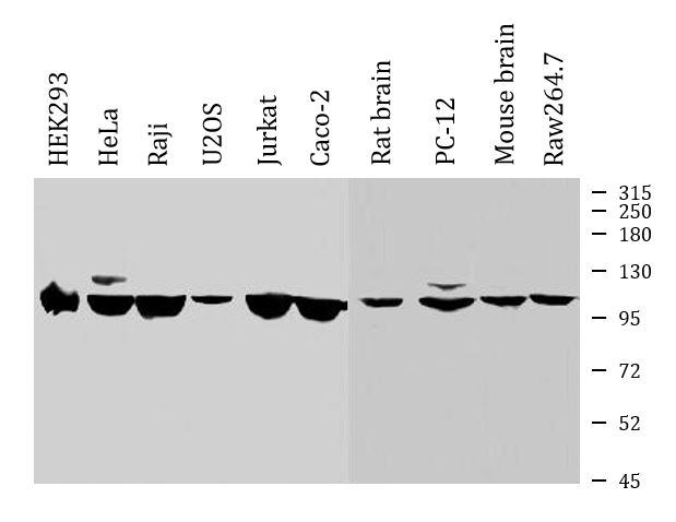
ARG43102 anti-Semaphorin 6A antibody WB image
Western blot: 50 µg of sample under reducing conditions. HEK293, HeLa, Raji, U2OS, Jurkat, Caco-2, Rat brain, PC-12, Mouse brain and Raw264.7 whole cell lysates stained with ARG43102 anti-Semaphorin 6A antibody at 0.25 µg/ml dilution, overnight at 4°C.
-

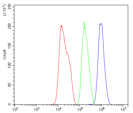
ARG43102 anti-Semaphorin 6A antibody FACS image
Flow Cytometry: HepG2 cells were blocked with 10% normal goat serum and then stained with ARG43102 anti-Semaphorin 6A antibody (blue) at 1 µg/10^6 cells for 30 min at 20°C, followed by incubation with DyLight®488 labelled secondary antibody. Isotype control antibody (green) was rabbit IgG (1 µg/10^6 cells) used under the same conditions. Unlabelled sample (red) was also used as a control.