ARG59542
anti-STUB1 / CHIP antibody
anti-STUB1 / CHIP antibody for Flow cytometry,ICC/IF,IHC-Formalin-fixed paraffin-embedded sections,Western blot and Human,Mouse,Rat
Overview
| Product Description | Rabbit Polyclonal antibody recognizes STUB1 / CHIP |
|---|---|
| Tested Reactivity | Hu, Ms, Rat |
| Tested Application | FACS, ICC/IF, IHC-P, WB |
| Host | Rabbit |
| Clonality | Polyclonal |
| Isotype | IgG |
| Target Name | STUB1 / CHIP |
| Antigen Species | Human |
| Immunogen | Recombinant protein corresponding to R51-Y303 of Human STUB1 / CHIP. |
| Conjugation | Un-conjugated |
| Alternate Names | NY-CO-7; Antigen NY-CO-7; UBOX1; EC 6.3.2.-; E3 ubiquitin-protein ligase CHIP; SDCCAG7; CHIP; STIP1 homology and U box-containing protein 1; HSPABP2; SCAR16; Carboxy terminus of Hsp70-interacting protein; CLL-associated antigen KW-8 |
Application Instructions
| Application Suggestion |
|
||||||||||
|---|---|---|---|---|---|---|---|---|---|---|---|
| Application Note | IHC-P: Antigen Retrieval: Heat mediation was performed in Citrate buffer (pH 6.0) for 20 min. * The dilutions indicate recommended starting dilutions and the optimal dilutions or concentrations should be determined by the scientist. |
Properties
| Form | Liquid |
|---|---|
| Purification | Affinity purification with immunogen. |
| Buffer | 0.9% NaCl, 0.2% Na2HPO4, 0.05% Sodium azide and 4% Trehalose. |
| Preservative | 0.05% Sodium azide |
| Stabilizer | 4% Trehalose |
| Concentration | 0.5 mg/ml |
| Storage Instruction | For continuous use, store undiluted antibody at 2-8°C for up to a week. For long-term storage, aliquot and store at -20°C or below. Storage in frost free freezers is not recommended. Avoid repeated freeze/thaw cycles. Suggest spin the vial prior to opening. The antibody solution should be gently mixed before use. |
| Note | For laboratory research only, not for drug, diagnostic or other use. |
Bioinformation
| Database Links |
Swiss-port # Q9UNE7 Human E3 ubiquitin-protein ligase CHIP Swiss-port # Q9WUD1 Mouse STIP1 homology and U box-containing protein 1 |
|---|---|
| Gene Symbol | STUB1 |
| Gene Full Name | STIP1 homology and U-box containing protein 1, E3 ubiquitin protein ligase |
| Background | This gene encodes a protein containing tetratricopeptide repeat and a U-box that functions as a ubiquitin ligase/cochaperone. The encoded protein binds to and ubiquitinates shock cognate 71 kDa protein (Hspa8) and DNA polymerase beta (Polb), among other targets. Mutations in this gene cause spinocerebellar ataxia, autosomal recessive 16. Alternative splicing results in multiple transcript variants. There is a pseudogene for this gene on chromosome 2. [provided by RefSeq, Jun 2014] |
| Function | E3 ubiquitin-protein ligase which targets misfolded chaperone substrates towards proteasomal degradation. Collaborates with ATXN3 in the degradation of misfolded chaperone substrates: ATXN3 restricting the length of ubiquitin chain attached to STUB1/CHIP substrates and preventing further chain extension. Ubiquitinates NOS1 in concert with Hsp70 and Hsp40. Modulates the activity of several chaperone complexes, including Hsp70, Hsc70 and Hsp90. Mediates transfer of non-canonical short ubiquitin chains to HSPA8 that have no effect on HSPA8 degradation. Mediates polyubiquitination of DNA polymerase beta (POLB) at 'Lys-41', 'Lys-61' and 'Lys-81', thereby playing a role in base-excision repair: catalyzes polyubiquitination by amplifying the HUWE1/ARF-BP1-dependent monoubiquitination and leading to POLB-degradation by the proteasome. Mediates polyubiquitination of CYP3A4. Ubiquitinates EPHA2 and may regulate the receptor stability and activity through proteasomal degradation. Negatively regulates the suppressive function of regulatory T-cells (Treg) during inflammation by mediating the ubiquitination and degradation of FOXP3 in a HSPA1A/B-dependent manner. [UniProt] |
| Cellular Localization | Cytoplasm. Nucleus. Note=Translocates to the nucleus in response to inflammatory signals in regulatory T-cells (Treg). [UniProt] |
| Calculated MW | 35 kDa |
| PTM | Monoubiquitinated at Lys-2 following cell stress by UBE2W, promoting the interaction with ATXN3 (By similarity). Auto-ubiquitinated; mediated by UBE2D1 and UBE2D2. [UniProt] |
Images (7) Click the Picture to Zoom In
-

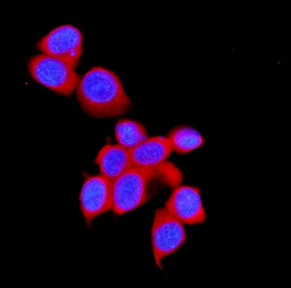
ARG59542 anti-STUB1 / CHIP antibody ICC/IF image
Immunofluorescence: MCF-7 cells were blocked with 10% goat serum and then stained with ARG59542 anti-STUB1 / CHIP antibody (red) at 2 µg/ml dilution, overnight at 4°C. DAPI (blue) for nuclear staining.
-
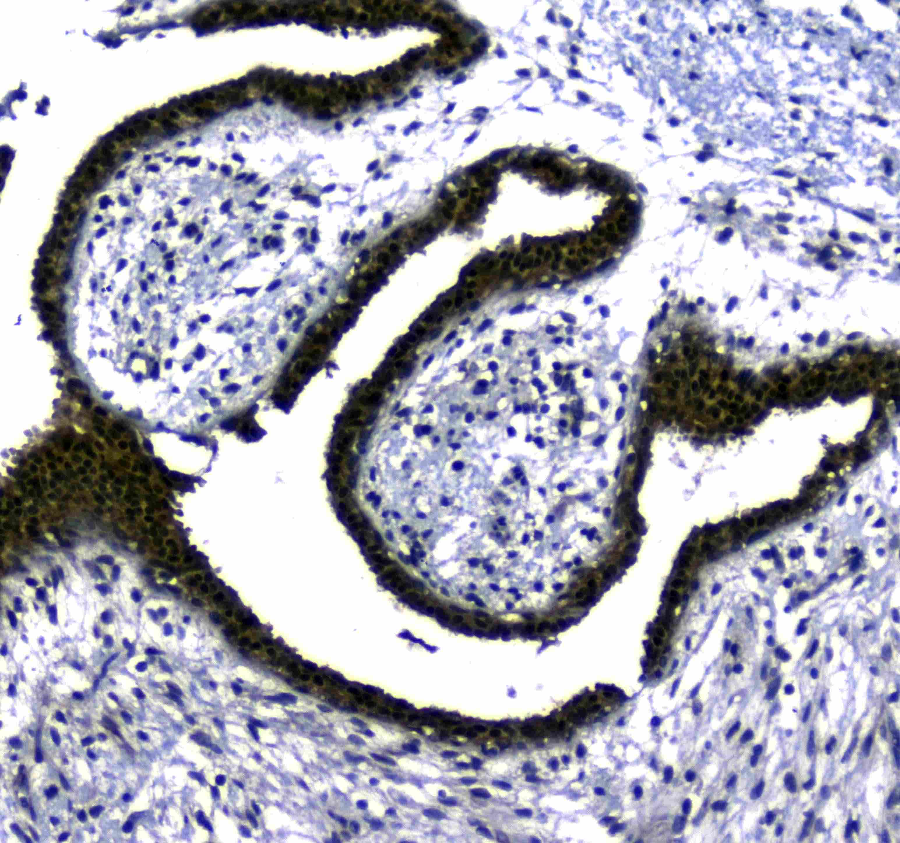
ARG59542 anti-STUB1 / CHIP antibody IHC-P image
Immunohistochemistry: Paraffin-embedded Human colon cancer tissue. Antigen Retrieval: Heat mediation was performed in Citrate buffer (pH 6.0, epitope retrieval solution) for 20 min. The tissue section was blocked with 10% goat serum. The tissue section was then stained with ARG59542 anti-STUB1 / CHIP antibody at 1 µg/ml, overnight at 4°C.
-
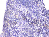
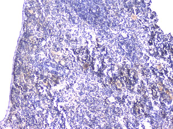
ARG59542 anti-STUB1 / CHIP antibody IHC-P image
Immunohistochemistry: Paraffin-embedded Mouse spleen tissue. Antigen Retrieval: Heat mediation was performed in Citrate buffer (pH 6.0) for 20 min. The tissue section was blocked with 10% goat serum. The tissue section was then stained with ARG59542 anti-STUB1 / CHIP antibody at 1 µg/ml dilution, overnight at 4°C.
-
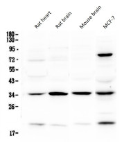
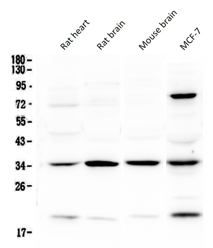
ARG59542 anti-STUB1 / CHIP antibody WB image
Western blot: 50 µg of samples under reducing conditions. Rat heart, Rat brain, Mouse brain and MCF-7 whole cell lysates stained with ARG59542 anti-STUB1 / CHIP antibody at 0.5 µg/ml, overnight at 4°C.
-
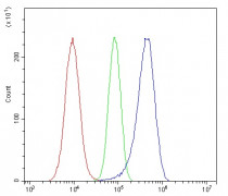
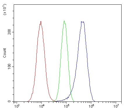
ARG59542 anti-STUB1 / CHIP antibody FACS image
Flow Cytometry: A549 cells were blocked with 10% normal goat serum and then stained with ARG59542 anti-STUB1 / CHIP antibody (blue) at 1 µg/10^6 cells for 30 min at 20°C, followed by incubation with DyLight®488 labelled secondary antibody. Isotype control antibody (green) was rabbit IgG (1 µg/10^6 cells) used under the same conditions. Unlabelled sample (red) was also used as a control.
-
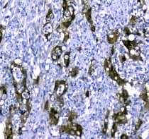
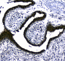
ARG59542 anti-STUB1 / CHIP antibody IHC-P image
Immunohistochemistry: Paraffin-embedded Human lung cancer tissue. Antigen Retrieval: Heat mediation was performed in Citrate buffer (pH 6.0, epitope retrieval solution) for 20 min. The tissue section was blocked with 10% goat serum. The tissue section was then stained with ARG59542 anti-STUB1 / CHIP antibody at 1 µg/ml, overnight at 4°C.
-
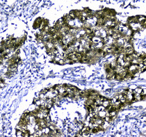
ARG59542 anti-STUB1 / CHIP antibody IHC-P image
Immunohistochemistry: Paraffin-embedded Human mammary cancer tissue. Antigen Retrieval: Heat mediation was performed in Citrate buffer (pH 6.0, epitope retrieval solution) for 20 min. The tissue section was blocked with 10% goat serum. The tissue section was then stained with ARG59542 anti-STUB1 / CHIP antibody at 1 µg/ml, overnight at 4°C.