ARG66700
anti-STAT6 phospho (Tyr641) antibody
anti-STAT6 phospho (Tyr641) antibody for IHC-Formalin-fixed paraffin-embedded sections,Western blot and Human,Mouse
Overview
| Product Description | Rabbit Polyclonal antibody recognizes STAT6 phospho (Tyr641) |
|---|---|
| Tested Reactivity | Hu, Ms |
| Predict Reactivity | Rat |
| Tested Application | IHC-P, WB |
| Host | Rabbit |
| Clonality | Polyclonal |
| Isotype | IgG |
| Target Name | STAT6 |
| Antigen Species | Human |
| Immunogen | Phosphospecific peptide around Tyr641 of Human STAT6. |
| Conjugation | Un-conjugated |
| Alternate Names | D12S1644; STAT6B; STAT6C; Signal transducer and activator of transcription 6; IL-4-STAT; IL-4 Stat |
Application Instructions
| Application Suggestion |
|
||||||
|---|---|---|---|---|---|---|---|
| Application Note | * The dilutions indicate recommended starting dilutions and the optimal dilutions or concentrations should be determined by the scientist. | ||||||
| Observed Size | ~ 95 kDa |
Properties
| Form | Liquid |
|---|---|
| Purification | Affinity purification with immunogen. |
| Buffer | PBS, 0.02% Sodium azide, 50% Glycerol and 0.5% BSA. |
| Preservative | 0.02% Sodium azide |
| Stabilizer | 50% Glycerol and 0.5% BSA |
| Concentration | 1 mg/ml |
| Storage Instruction | For continuous use, store undiluted antibody at 2-8°C for up to a week. For long-term storage, aliquot and store at -20°C. Storage in frost free freezers is not recommended. Avoid repeated freeze/thaw cycles. Suggest spin the vial prior to opening. The antibody solution should be gently mixed before use. |
| Note | For laboratory research only, not for drug, diagnostic or other use. |
Bioinformation
| Database Links |
Swiss-port # P42226 Human Signal transducer and activator of transcription 6 Swiss-port # P52633 Mouse Signal transducer and transcription activator 6 |
|---|---|
| Gene Symbol | STAT6 |
| Gene Full Name | signal transducer and activator of transcription 6, interleukin-4 induced |
| Background | The protein encoded by this gene is a member of the STAT family of transcription factors. In response to cytokines and growth factors, STAT family members are phosphorylated by the receptor associated kinases, and then form homo- or heterodimers that translocate to the cell nucleus where they act as transcription activators. This protein plays a central role in exerting IL4 mediated biological responses. It is found to induce the expression of BCL2L1/BCL-X(L), which is responsible for the anti-apoptotic activity of IL4. Knockout studies in mice suggested the roles of this gene in differentiation of T helper 2 (Th2) cells, expression of cell surface markers, and class switch of immunoglobulins. Alternative splicing results in multiple transcript variants.[provided by RefSeq, May 2010] |
| Function | Carries out a dual function: signal transduction and activation of transcription. Involved in IL4/interleukin-4- and IL3/interleukin-3-mediated signaling. [UniProt] |
| Cellular Localization | Cytoplasm. Nucleus. Note=Translocated into the nucleus in response to phosphorylation. [UniProt] |
| Calculated MW | 94 kDa |
| PTM | Tyrosine phosphorylated following stimulation by IL4/interleukin-4 and IL3/interleukin-3 (By similarity). Dephosphorylation on tyrosine residues by PTPN2 negatively regulates the IL4/interleukin-4 mediated signaling. [UniProt] |
Images (4) Click the Picture to Zoom In
-

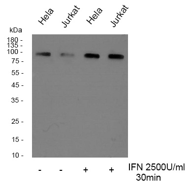
ARG66700 anti-STAT6 phospho (Tyr641) antibody WB image
Western blot: HeLa and Jurkat cells were untreated or treated with IFN 2500U/ml for 30 minutes, overnight at 4°C. Cell lysates were stained with ARG66700 anti-STAT6 phospho (Tyr641) antibody.
-
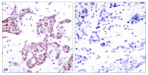
ARG66700 anti-STAT6 phospho (Tyr641) antibody IHC-P image
Immunohistochemistry: Paraffin-embedded Human breast carcinoma tissue stained with ARG66700 anti-STAT6 phospho (Tyr641) antibody. The picture on the right is blocked with the phospho peptide.
-

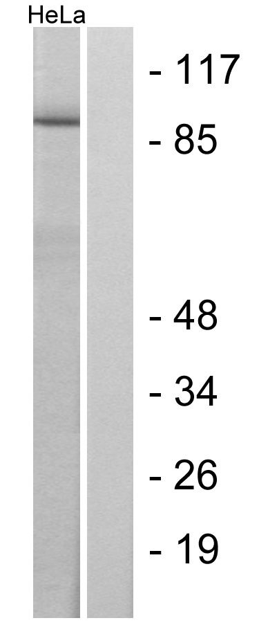
ARG66700 anti-STAT6 phospho (Tyr641) antibody WB image
Western blot: HeLa cells treated with IL4. Cell lysates were stained with ARG66700 anti-STAT6 phospho (Tyr641) antibody. The lane on the right is blocked with the phospho peptide.
-
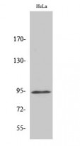
ARG66700 anti-STAT6 phospho (Tyr641) antibody WB image
Western blot: HeLa cell lysate stained with ARG66700 anti-STAT6 phospho (Tyr641) antibody.