ARG65593
anti-STAT5A antibody
anti-STAT5A antibody for IHC-Formalin-fixed paraffin-embedded sections,Western blot and Human
Gene Regulation antibody; Signaling Transduction antibody
Overview
| Product Description | Rabbit Polyclonal antibody recognizes STAT5A |
|---|---|
| Tested Reactivity | Hu |
| Tested Application | IHC-P, WB |
| Host | Rabbit |
| Clonality | Polyclonal |
| Isotype | IgG |
| Target Name | STAT5A |
| Antigen Species | Human |
| Immunogen | Synthetic peptide of human STAT5A |
| Conjugation | Un-conjugated |
| Alternate Names | Signal transducer and activator of transcription 5A; STAT5; MGF |
Application Instructions
| Application Suggestion |
|
||||||
|---|---|---|---|---|---|---|---|
| Application Note | * The dilutions indicate recommended starting dilutions and the optimal dilutions or concentrations should be determined by the scientist. |
Properties
| Form | Liquid |
|---|---|
| Purification | Purified by antigen-affinity chromatography. |
| Buffer | PBS (pH 7.4), 0.05% Sodium azide and 40% Glycerol |
| Preservative | 0.05% Sodium azide |
| Stabilizer | 40% Glycerol |
| Concentration | 1 mg/ml |
| Storage Instruction | For continuous use, store undiluted antibody at 2-8°C for up to a week. For long-term storage, aliquot and store at -20°C. Storage in frost free freezers is not recommended. Avoid repeated freeze/thaw cycles. Suggest spin the vial prior to opening. The antibody solution should be gently mixed before use. |
| Note | For laboratory research only, not for drug, diagnostic or other use. |
Bioinformation
| Database Links |
Swiss-port # P42229 Human Signal transducer and activator of transcription 5A |
|---|---|
| Gene Symbol | STAT5A |
| Gene Full Name | signal transducer and activator of transcription 5A |
| Background | The protein encoded by this gene is a member of the STAT family of transcription factors. In response to cytokines and growth factors, STAT family members are phosphorylated by the receptor associated kinases, and then form homo- or heterodimers that translocate to the cell nucleus where they act as transcription activators. This protein is activated by, and mediates the responses of many cell ligands, such as IL2, IL3, IL7 GM-CSF, erythropoietin, thrombopoietin, and different growth hormones. Activation of this protein in myeloma and lymphoma associated with a TEL/JAK2 gene fusion is independent of cell stimulus and has been shown to be essential for the tumorigenesis. The mouse counterpart of this gene is found to induce the expression of BCL2L1/BCL-X(L), which suggests the antiapoptotic function of this gene in cells. |
| Function | Carries out a dual function: signal transduction and activation of transcription. Mediates cellular responses to the cytokine KITLG/SCF and other growth factors. Mediates cellular responses to ERBB4. May mediate cellular responses to activated FGFR1, FGFR2, FGFR3 and FGFR4. Binds to the GAS element and activates PRL-induced transcription. Regulates the expression of milk proteins during lactation. [UniProt] |
| Highlight | Related Antibody Duos and Panels: ARG30214 Phospho STAT5A Antibody Duo (Total, pY694) Related products: STAT5A antibodies; STAT5A Duos / Panels; Anti-Rabbit IgG secondary antibodies; |
| Research Area | Gene Regulation antibody; Signaling Transduction antibody |
| Calculated MW | 91 kDa |
| PTM | Tyrosine phosphorylated in response to KITLG/SCF, IL2, IL3, IL7, IL15, CSF2/GMCSF, GH1, PRL, EPO and THPO. Activated KIT promotes phosphorylation on tyrosine residues and subsequent translocation to the nucleus. Tyrosine phosphorylated in response to constitutively activated FGFR1, FGFR2, FGFR3 and FGFR4. Tyrosine phosphorylation is required for DNA-binding activity and dimerization. Serine phosphorylation is also required for maximal transcriptional activity (By similarity). Tyrosine phosphorylated in response to signaling via activated FLT3; wild-type FLT3 results in much weaker phosphorylation than constitutively activated mutant FLT3. Alternatively, can be phosphorylated by JAK2 at Tyr-694. Dephosphorylation on tyrosine residues by PTPN2 negatively regulates prolactin signaling pathway. ISGylated. |
Images (2) Click the Picture to Zoom In
-
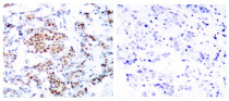
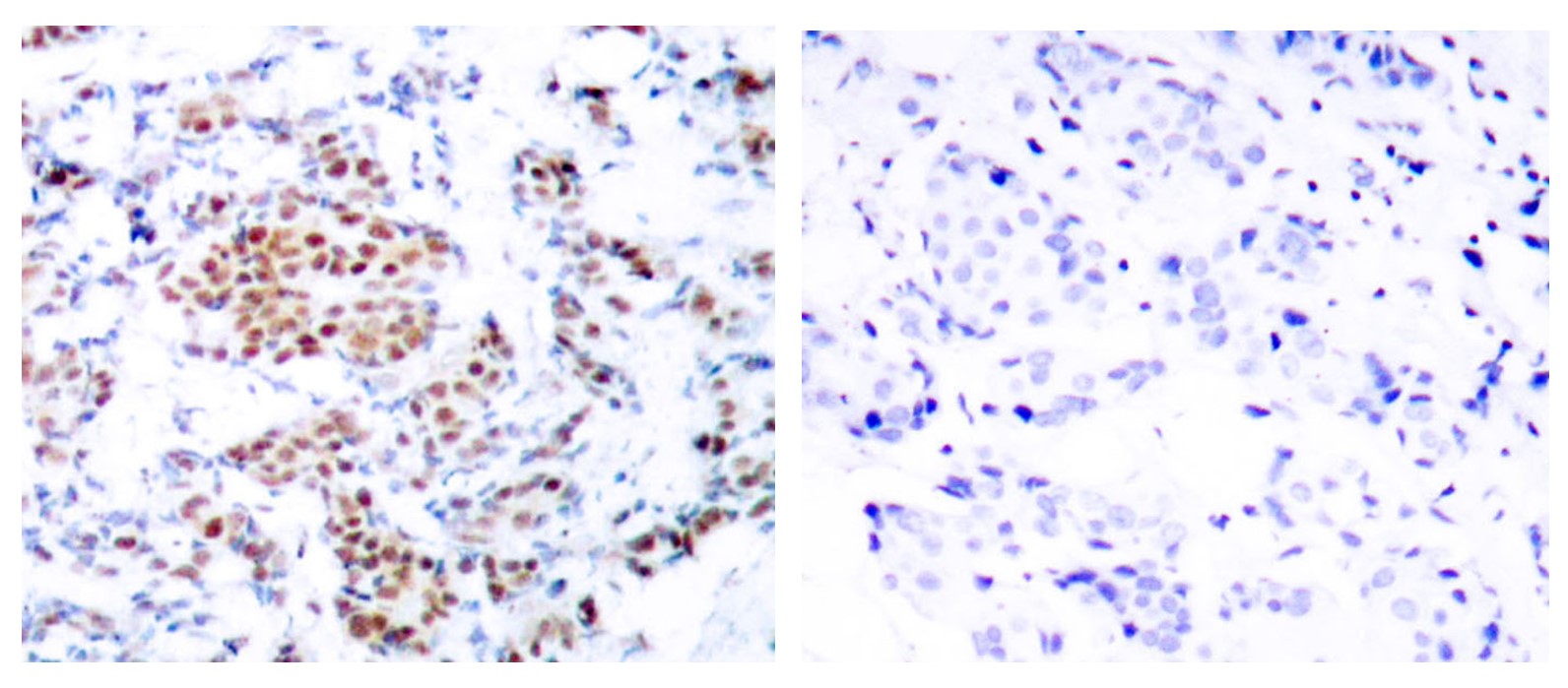
ARG65593 anti-STAT5A antibody IHC-P image
Immunohistochemistry: Paraffin-embedded Human breast carcinoma tissue stained with ARG65593 anti-STAT5A antibody at 1:50 dilution, or the same antibody preincubated with synthetic peptide (right). (Original magnification: ×200).
-
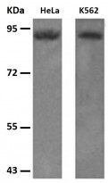
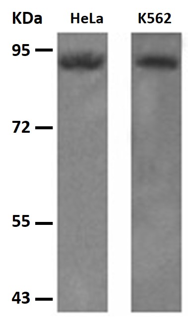
ARG65593 anti-STAT5A antibody WB image
Western blot: 30 μg of HeLa and K562 cell lysates stained with ARG65593 anti-STAT5A antibody at 1:500 dilution. Exposure time: 1 minute.