ARG57263
anti-STAT3 phospho (Tyr705) antibody [RM261]
anti-STAT3 phospho (Tyr705) antibody [RM261] for IHC-Formalin-fixed paraffin-embedded sections,Western blot and Human
Overview
| Product Description | Rabbit Monoclonal antibody [RM261] recognizes STAT3 phospho (Tyr705) |
|---|---|
| Tested Reactivity | Hu |
| Predict Reactivity | Ms, Rat |
| Tested Application | IHC-P, WB |
| Specificity | This antibody reacts to Human Stat3 only when phosphorylated at Tyr705. There is no cross-reactivity to stat3 without phosphorylation at Tyr705. This antibody may also react to Mouse or Rat Phospho-Stat3 (Tyr705), as predicted by immunogen homology. |
| Host | Rabbit |
| Clonality | Monoclonal |
| Clone | RM261 |
| Isotype | IgG |
| Target Name | STAT3 |
| Antigen Species | Human |
| Immunogen | Phospho-peptide around Tyr705 of Human STAT3. |
| Conjugation | Un-conjugated |
| Alternate Names | ADMIO; APRF; HIES; Acute-phase response factor; Signal transducer and activator of transcription 3 |
Application Instructions
| Application Suggestion |
|
||||||
|---|---|---|---|---|---|---|---|
| Application Note | * The dilutions indicate recommended starting dilutions and the optimal dilutions or concentrations should be determined by the scientist. |
Properties
| Form | Liquid |
|---|---|
| Purification | Purification with Protein A. |
| Buffer | PBS, 0.09% Sodium azide, 50% Glycerol and 1% BSA. |
| Preservative | 0.09% Sodium azide |
| Stabilizer | 50% Glycerol and 1% BSA |
| Storage Instruction | For continuous use, store undiluted antibody at 2-8°C for up to a week. For long-term storage, aliquot and store at -20°C. Storage in frost free freezers is not recommended. Avoid repeated freeze/thaw cycles. Suggest spin the vial prior to opening. The antibody solution should be gently mixed before use. |
| Note | For laboratory research only, not for drug, diagnostic or other use. |
Bioinformation
| Database Links |
Swiss-port # P40763 Human Signal transducer and activator of transcription 3 |
|---|---|
| Gene Symbol | STAT3 |
| Gene Full Name | signal transducer and activator of transcription 3 (acute-phase response factor) |
| Background | The protein encoded by this gene is a member of the STAT protein family. In response to cytokines and growth factors, STAT family members are phosphorylated by the receptor associated kinases, and then form homo- or heterodimers that translocate to the cell nucleus where they act as transcription activators. This protein is activated through phosphorylation in response to various cytokines and growth factors including IFNs, EGF, IL5, IL6, HGF, LIF and BMP2. This protein mediates the expression of a variety of genes in response to cell stimuli, and thus plays a key role in many cellular processes such as cell growth and apoptosis. The small GTPase Rac1 has been shown to bind and regulate the activity of this protein. PIAS3 protein is a specific inhibitor of this protein. Mutations in this gene are associated with infantile-onset multisystem autoimmune disease and hyper-immunoglobulin E syndrome. Alternative splicing results in multiple transcript variants encoding distinct isoforms. [provided by RefSeq, Sep 2015] |
| Function | Signal transducer and transcription activator that mediates cellular responses to interleukins, KITLG/SCF and other growth factors. May mediate cellular responses to activated FGFR1, FGFR2, FGFR3 and FGFR4. Binds to the interleukin-6 (IL-6)-responsive elements identified in the promoters of various acute-phase protein genes. Activated by IL31 through IL31RA. Cytoplasmic STAT3 represses macroautophagy by inhibiting EIF2AK2/PKR activity. Plays an important role in host defense in methicillin-resistant S.aureus lung infection by regulating the expression of the antimicrobial lectin REG3G (By similarity). [UniProt] |
| Highlight | Related products: STAT3 antibodies; Anti-Rabbit IgG secondary antibodies; Related news: LKB1 deficiency in T cells promotes gut tumors |
| Calculated MW | 88 kDa |
| PTM | Tyrosine phosphorylated upon stimulation with EGF. Tyrosine phosphorylated in response to constitutively activated FGFR1, FGFR2, FGFR3 and FGFR4 (By similarity). Activated through tyrosine phosphorylation by BMX. Tyrosine phosphorylated in response to IL6, IL11, LIF, CNTF, KITLG/SCF, CSF1, EGF, PDGF, IFN-alpha, LEP and OSM. Activated KIT promotes phosphorylation on tyrosine residues and subsequent translocation to the nucleus. Phosphorylated on serine upon DNA damage, probably by ATM or ATR. Serine phosphorylation is important for the formation of stable DNA-binding STAT3 homodimers and maximal transcriptional activity. ARL2BP may participate in keeping the phosphorylated state of STAT3 within the nucleus. Upon LPS challenge, phosphorylated within the nucleus by IRAK1. Upon erythropoietin treatment, phosphorylated on Ser-727 by RPS6KA5. Phosphorylation at Tyr-705 by PTK6 or FER leads to an increase of its transcriptional activity. Dephosphorylation on tyrosine residues by PTPN2 negatively regulates IL6/interleukin-6 signaling. Acetylated on lysine residues by CREBBP. Deacetylation by LOXL3 leads to disrupt STAT3 dimerization and inhibit STAT3 transcription activity (PubMed:28065600). Oxidation of lysine residues to allysine on STAT3 preferentially takes place on lysine residues that are acetylated (PubMed:28065600). Some lysine residues are oxidized to allysine by LOXL3, leading to disrupt STAT3 dimerization and inhibit STAT3 transcription activity (PubMed:28065600). Oxidation of lysine residues to allysine on STAT3 preferentially takes place on lysine residues that are acetylated (PubMed:28065600). |
Images (2) Click the Picture to Zoom In
-
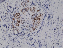
ARG57263 anti-STAT3 phospho (Tyr705) antibody [RM261] IHC-P image
Immunohistochemistry: Formalin-fixed and paraffin-embedded Human breast cancer tissue sections stained with ARG57263 anti-STAT3 phospho (Tyr705) antibody [RM261] at 1:10000 dilution.
-
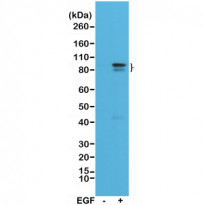
ARG57263 anti-STAT3 phospho (Tyr705) antibody [RM261] WB image
Western blot: A431 cell lysates, nontreated (-) or treated (+) with EGF, stained with ARG57263 anti-STAT3 phospho (Tyr705) antibody [RM261] at 1:1000 dilution, showed Phospho-Stat3 (Tyr705) in EGF-treated A431 cells.


