ARG63340
anti-STAT3 antibody
anti-STAT3 antibody for Flow cytometry,ICC/IF,IHC-Formalin-fixed paraffin-embedded sections,Western blot and Human,Mouse
Cancer antibody; Cell Biology and Cellular Response antibody; Developmental Biology antibody; Gene Regulation antibody; Signaling Transduction antibody
Overview
| Product Description | Goat Polyclonal antibody recognizes STAT3 |
|---|---|
| Tested Reactivity | Hu, Ms |
| Predict Reactivity | Cow, Rat, Dog, Pig |
| Tested Application | FACS, ICC/IF, IHC-P, WB |
| Specificity | This antibody is expected to recognise isoforms 1 and 2 (as represented by NP_644805.1 and NP_003141.2 respectively) |
| Host | Goat |
| Clonality | Polyclonal |
| Isotype | IgG |
| Target Name | STAT3 |
| Antigen Species | Human |
| Immunogen | DMELTSECATSPM |
| Conjugation | Un-conjugated |
| Alternate Names | ADMIO; APRF; HIES; Acute-phase response factor; Signal transducer and activator of transcription 3 |
Application Instructions
| Application Suggestion |
|
||||||||||
|---|---|---|---|---|---|---|---|---|---|---|---|
| Application Note | WB: Recommend incubate at RT for 1h. IHC-P: Antigen Retrieval: Microwaved tissue section in Citrate buffer (pH 6.0). * The dilutions indicate recommended starting dilutions and the optimal dilutions or concentrations should be determined by the scientist. |
Properties
| Form | Liquid |
|---|---|
| Purification | Purified from goat serum by antigen affinity chromatography. |
| Buffer | Tris saline (pH 7.3), 0.02% Sodium azide and 0.5% BSA. |
| Preservative | 0.02% Sodium azide |
| Stabilizer | 0.5% BSA |
| Concentration | 0.5 mg/ml |
| Storage Instruction | For continuous use, store undiluted antibody at 2-8°C for up to a week. For long-term storage, aliquot and store at -20°C or below. Storage in frost free freezers is not recommended. Avoid repeated freeze/thaw cycles. Suggest spin the vial prior to opening. The antibody solution should be gently mixed before use. |
| Note | For laboratory research only, not for drug, diagnostic or other use. |
Bioinformation
| Database Links |
Swiss-port # P40763 Human Signal transducer and activator of transcription 3 Swiss-port # P42227 Mouse Signal transducer and activator of transcription 3 |
|---|---|
| Gene Symbol | Stat3 |
| Gene Full Name | signal transducer and activator of transcription 3 (acute-phase response factor) |
| Background | The protein encoded by this gene is a member of the STAT protein family. In response to cytokines and growth factors, STAT family members are phosphorylated by the receptor associated kinases, and then form homo- or heterodimers that translocate to the cell nucleus where they act as transcription activators. This protein is activated through phosphorylation in response to various cytokines and growth factors including IFNs, EGF, IL5, IL6, HGF, LIF and BMP2. This protein mediates the expression of a variety of genes in response to cell stimuli, and thus plays a key role in many cellular processes such as cell growth and apoptosis. The small GTPase Rac1 has been shown to bind and regulate the activity of this protein. PIAS3 protein is a specific inhibitor of this protein. Three alternatively spliced transcript variants encoding distinct isoforms have been described. [provided by RefSeq, Jul 2008] |
| Research Area | Cancer antibody; Cell Biology and Cellular Response antibody; Developmental Biology antibody; Gene Regulation antibody; Signaling Transduction antibody |
| Calculated MW | 88 kDa |
| PTM | Tyrosine phosphorylated upon stimulation with EGF. Tyrosine phosphorylated in response to constitutively activated FGFR1, FGFR2, FGFR3 and FGFR4 (By similarity). Activated through tyrosine phosphorylation by BMX. Tyrosine phosphorylated in response to IL6, IL11, LIF, CNTF, KITLG/SCF, CSF1, EGF, PDGF, IFN-alpha, LEP and OSM. Activated KIT promotes phosphorylation on tyrosine residues and subsequent translocation to the nucleus. Phosphorylated on serine upon DNA damage, probably by ATM or ATR. Serine phosphorylation is important for the formation of stable DNA-binding STAT3 homodimers and maximal transcriptional activity. ARL2BP may participate in keeping the phosphorylated state of STAT3 within the nucleus. Upon LPS challenge, phosphorylated within the nucleus by IRAK1. Upon erythropoietin treatment, phosphorylated on Ser-727 by RPS6KA5. Phosphorylation at Tyr-705 by PTK6 or FER leads to an increase of its transcriptional activity. Dephosphorylation on tyrosine residues by PTPN2 negatively regulates IL6/interleukin-6 signaling. Acetylated on lysine residues by CREBBP. Deacetylation by LOXL3 leads to disrupt STAT3 dimerization and inhibit STAT3 transcription activity (PubMed:28065600). Oxidation of lysine residues to allysine on STAT3 preferentially takes place on lysine residues that are acetylated (PubMed:28065600). Some lysine residues are oxidized to allysine by LOXL3, leading to disrupt STAT3 dimerization and inhibit STAT3 transcription activity (PubMed:28065600). Oxidation of lysine residues to allysine on STAT3 preferentially takes place on lysine residues that are acetylated (PubMed:28065600). |
Images (5) Click the Picture to Zoom In
-

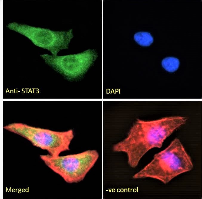
ARG63340 anti-STAT3 antibody ICC/IF image
Immunofluorescence: Paraformaldehyde fixed HeLa cells permeabilized with 0.15% Triton. Cells were stained with ARG63340 anti-STAT3 antibody (green) at 10 µg/ml dilution for 1 hour. DAPI (blue) for nuclear staining. Phalloidin (red) for Actin filaments staining. Negative control: Unimmunized goat IgG (green) at 10 µg/ml dilution.
-
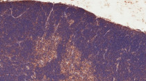
ARG63340 anti-STAT3 antibody IHC-P image
Immunohistochemistry: Paraffin-embedded Mouse thymus tissue. Antigen Retrieval: Microwaved tissue section in Citrate buffer (pH 6.0). The tissue section was stained with ARG63340 anti-STAT3 antibody at 4 µg/ml dilution followed by HRP-staining.
-
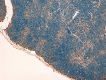
ARG63340 anti-STAT3 antibody IHC-P image
Immunohistochemistry: paraffin embedded Mouse Thymus. (Steamed antigen retrieval with Tris/EDTA buffer pH 9) stained with ARG63340 anti-STAT3 (isoform 1 and 2) antibody at 4 µg/ml dilution followed by HRP-staining
-
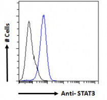
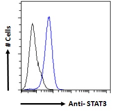
ARG63340 anti-STAT3 antibody FACS image
Flow Cytometry: Paraformaldehyde-fixed A431 cells permeabilized with 0.5% Triton. Cells were stained with ARG63340 anti-STAT3 antibody (blue line) at 10 µg/ml dilution for 1 hour, followed by incubation with Alexa FluorR 488 labelled secondary antibody. IgG control: Unimmunized goat IgG (black line).
-
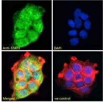
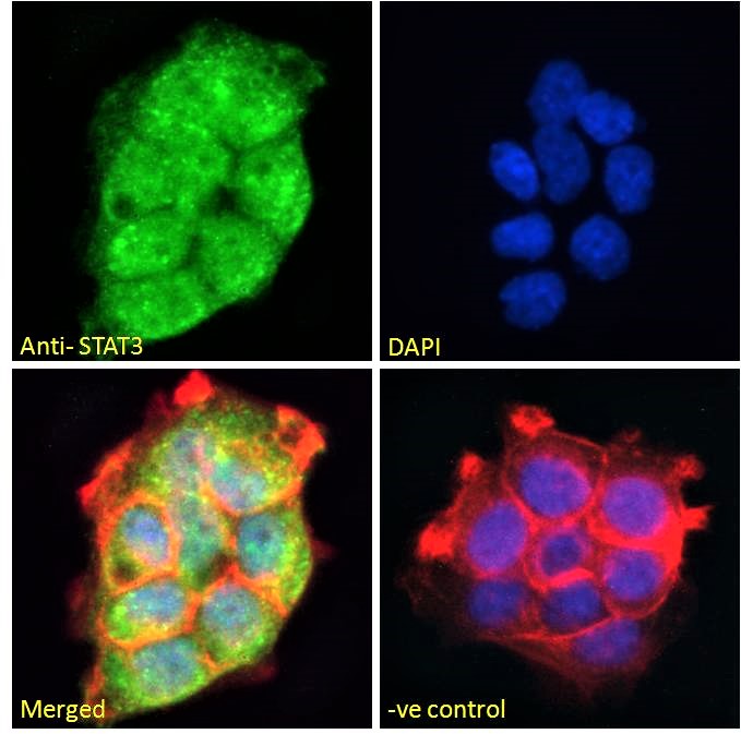
ARG63340 anti-STAT3 antibody ICC/IF image
Immunofluorescence: Paraformaldehyde fixed A431 cells permeabilized with 0.15% Triton. Cells were stained with ARG63340 anti-STAT3 antibody (green) at 10 µg/ml dilution for 1 hour. DAPI (blue) for nuclear staining. Phalloidin (red) for Actin filaments staining. Negative control: Unimmunized goat IgG (green) at 10 µg/ml dilution.