ARG57136
anti-ST13 / Hip antibody [5C6]
anti-ST13 / Hip antibody [5C6] for Flow cytometry,ICC/IF,Western blot and Human
Overview
| Product Description | Mouse Monoclonal antibody [5C6] recognizes ST13 / Hip |
|---|---|
| Tested Reactivity | Hu |
| Tested Application | FACS, ICC/IF, WB |
| Host | Mouse |
| Clonality | Monoclonal |
| Clone | 5C6 |
| Isotype | IgG2a, kappa |
| Target Name | ST13 / Hip |
| Antigen Species | Human |
| Immunogen | Recombinant fragment around aa. 1-369 of Human ST13 / Hip |
| Conjugation | Un-conjugated |
| Alternate Names | HIP; SNC6; Putative tumor suppressor ST13; Progesterone receptor-associated p48 protein; Suppression of tumorigenicity 13 protein; HSPABP; Hsc70-interacting protein; HSPABP1; Protein FAM10A1; Hip; AAG2; HOP; P48; FAM10A1; PRO0786; Renal carcinoma antigen NY-REN-33; Aging-associated protein 2; FAM10A4 |
Application Instructions
| Application Suggestion |
|
||||||||
|---|---|---|---|---|---|---|---|---|---|
| Application Note | * The dilutions indicate recommended starting dilutions and the optimal dilutions or concentrations should be determined by the scientist. |
Properties
| Form | Liquid |
|---|---|
| Purification | Purification with Protein A. |
| Buffer | PBS (pH 7.4), 0.02% Sodium azide and 10% Glycerol. |
| Preservative | 0.02% Sodium azide |
| Stabilizer | 10% Glycerol |
| Concentration | 1 mg/ml |
| Storage Instruction | For continuous use, store undiluted antibody at 2-8°C for up to a week. For long-term storage, aliquot and store at -20°C. Storage in frost free freezers is not recommended. Avoid repeated freeze/thaw cycles. Suggest spin the vial prior to opening. The antibody solution should be gently mixed before use. |
| Note | For laboratory research only, not for drug, diagnostic or other use. |
Bioinformation
| Database Links | |
|---|---|
| Gene Symbol | ST13 |
| Gene Full Name | suppression of tumorigenicity 13 (colon carcinoma) (Hsp70 interacting protein) |
| Background | The protein encoded by this gene is an adaptor protein that mediates the association of the heat shock proteins HSP70 and HSP90. This protein has been shown to be involved in the assembly process of glucocorticoid receptor, which requires the assistance of multiple molecular chaperones. The expression of this gene is reported to be downregulated in colorectal carcinoma tissue suggesting that it is a candidate tumor suppressor gene. Alternative splicing results in multiple transcript variants encoding different isoforms. [provided by RefSeq, Jun 2013] |
| Function | One HIP oligomer binds the ATPase domains of at least two HSC70 molecules dependent on activation of the HSC70 ATPase by HSP40. Stabilizes the ADP state of HSC70 that has a high affinity for substrate protein. Through its own chaperone activity, it may contribute to the interaction of HSC70 with various target proteins (By similarity). [UniProt] |
| Calculated MW | 41 kDa |
Images (6) Click the Picture to Zoom In
-
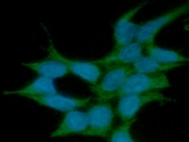
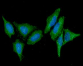
ARG57136 anti-ST13 / Hip antibody [5C6] ICC/IF image
Immunofluorescence: 293T cells line stained with ARG57136 anti-ST13 / Hip antibody [5C6] at 1:100 (Green).
DAPI (Blue) for nucleus staining.
-

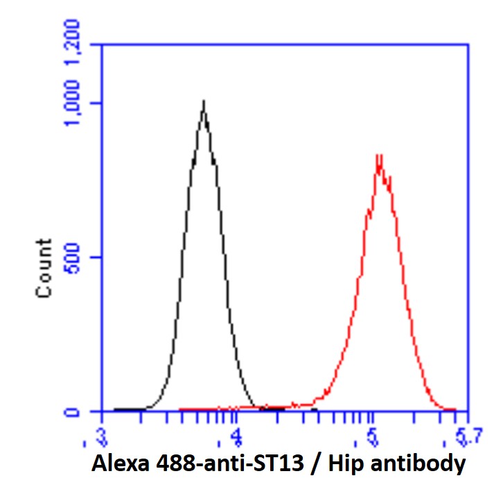
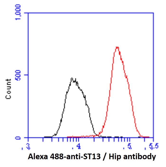
ARG57136 anti-ST13 / Hip antibody [5C6] FACS image
Flow Cytometry: 293T cell line stained with ARG57136 anti-ST13 / Hip antibody [5C6] at 2-5 µg for 1x10^6 cells (red line). Secondary antibody: Goat anti-Mouse IgG Alexa fluor 488 conjugate. Isotype control antibody: Mouse IgG (black line).
-
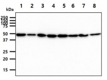
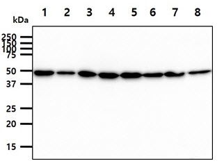
ARG57136 anti-ST13 / Hip antibody [5C6] WB image
Western blot: 40 µg of 1) 293T, 2) HepG2, 3) SW480, 4) Jurkat, 5) K562, 6) LnCap, 7) HeLa, and 8) PC3 cell lysates stained with ARG57136 anti-ST13 / Hip antibody [5C6] at 1:1000.
-
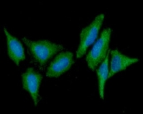
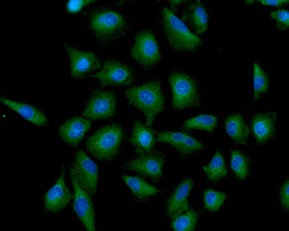
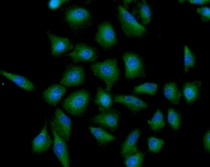
ARG57136 anti-ST13 / Hip antibody [5C6] ICC/IF image
Immunofluorescence: HeLa cells line stained with ARG57136 anti-ST13 / Hip antibody [5C6] at 1:100 (Green).
DAPI (Blue) for nucleus staining.
-
ARG57136 anti-ST13 / Hip antibody [5C6] ICC/IF image
Immunofluorescence: A549 cells line stained with ARG57136 anti-ST13 / Hip antibody [5C6] at 1:100 (Green).
DAPI (Blue) for nucleus staining.
-
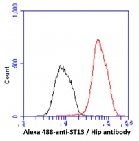
ARG57136 anti-ST13 / Hip antibody [5C6] FACS image
Flow Cytometry: Hep3B cell line stained with ARG57136 anti-ST13 / Hip antibody [5C6] at 2-5 µg for 1x10^6 cells (red line). Secondary antibody: Goat anti-Mouse IgG Alexa fluor 488 conjugate. Isotype control antibody: Mouse IgG (black line).