ARG63975
anti-SLIT2 antibody
anti-SLIT2 antibody for IHC-Formalin-fixed paraffin-embedded sections and Human
Neuroscience antibody
Overview
| Product Description | Goat Polyclonal antibody recognizes SLIT2 |
|---|---|
| Tested Reactivity | Hu |
| Predict Reactivity | Ms, Rat, Dog |
| Tested Application | IHC-P |
| Host | Goat |
| Clonality | Polyclonal |
| Isotype | IgG |
| Target Name | SLIT2 |
| Antigen Species | Human |
| Immunogen | DDCQDNKCKNGAH |
| Conjugation | Un-conjugated |
| Alternate Names | Slit-2; Slit homolog 2 protein; SLIL3 |
Application Instructions
| Application Suggestion |
|
||||
|---|---|---|---|---|---|
| Application Note | IHC-P: Antigen Retrieval: Steam tissue section in Tris/EDTA buffer (pH 9.0). * The dilutions indicate recommended starting dilutions and the optimal dilutions or concentrations should be determined by the scientist. |
Properties
| Form | Liquid |
|---|---|
| Purification | Purified from goat serum by antigen affinity chromatography. |
| Buffer | Tris saline (pH 7.3), 0.02% Sodium azide and 0.5% BSA. |
| Preservative | 0.02% Sodium azide |
| Stabilizer | 0.5% BSA |
| Concentration | 0.5 mg/ml |
| Storage Instruction | For continuous use, store undiluted antibody at 2-8°C for up to a week. For long-term storage, aliquot and store at -20°C or below. Storage in frost free freezers is not recommended. Avoid repeated freeze/thaw cycles. Suggest spin the vial prior to opening. The antibody solution should be gently mixed before use. |
| Note | For laboratory research only, not for drug, diagnostic or other use. |
Bioinformation
| Database Links | |
|---|---|
| Gene Symbol | SLIT2 |
| Gene Full Name | slit homolog 2 (Drosophila) |
| Background | This gene encodes a member of the slit family of secreted glycoproteins, which are ligands for the Robo family of immunoglobulin receptors. Slit proteins play highly conserved roles in axon guidance and neuronal migration and may also have functions during other cell migration processes including leukocyte migration. Members of the slit family are characterized by an N-terminal signal peptide, four leucine-rich repeats, nine epidermal growth factor repeats, and a C-terminal cysteine knot. Proteolytic processing of this protein gives rise to an N-terminal fragment that contains the four leucine-rich repeats and five epidermal growth factor repeats and a C-terminal fragment that contains four epidermal growth factor repeats and the cysteine knot. Both full length and cleaved proteins are secreted extracellularly and can function in axon repulsion as well as other specific processes. Alternative splicing results in multiple transcript variants. [provided by RefSeq, Sep 2015] |
| Function | Thought to act as molecular guidance cue in cellular migration, and function appears to be mediated by interaction with roundabout homolog receptors. During neural development involved in axonal navigation at the ventral midline of the neural tube and projection of axons to different regions. SLIT1 and SLIT2 seem to be essential for midline guidance in the forebrain by acting as repulsive signal preventing inappropriate midline crossing by axons projecting from the olfactory bulb. In spinal chord development may play a role in guiding commissural axons once they reached the floor plate by modulating the response to netrin. In vitro, silences the attractive effect of NTN1 but not its growth-stimulatory effect and silencing requires the formation of a ROBO1-DCC complex. May be implicated in spinal chord midline post-crossing axon repulsion. In vitro, only commissural axons that crossed the midline responded to SLIT2. In the developing visual system appears to function as repellent for retinal ganglion axons by providing a repulsion that directs these axons along their appropriate paths prior to, and after passage through, the optic chiasm. In vitro, collapses and repels retinal ganglion cell growth cones. Seems to play a role in branching and arborization of CNS sensory axons, and in neuronal cell migration. In vitro, Slit homolog 2 protein N-product, but not Slit homolog 2 protein C-product, repels olfactory bulb (OB) but not dorsal root ganglia (DRG) axons, induces OB growth cones collapse and induces branching of DRG axons. Seems to be involved in regulating leukocyte migration. [UniProt] |
| Research Area | Neuroscience antibody |
| Calculated MW | 170 kDa |
Images (1) Click the Picture to Zoom In
-
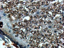
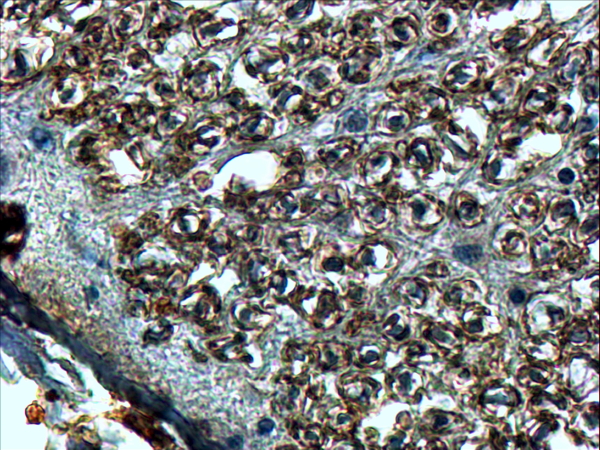
ARG63975 anti-SLIT2 antibody IHC-P image
Immunohistochemistry: paraffin embedded Human Spinal Cord. (Steamed antigen retrieval with Tris/EDTA buffer pH 9) stained with ARG63975 anti-SLIT2 antibody at 4 µg/ml dilution followed by HRP-staining.