ARG63447
anti-SHP2 antibody
anti-SHP2 antibody for Flow cytometry,ICC/IF,IHC-Formalin-fixed paraffin-embedded sections,Western blot and Human
Neuroscience antibody; Signaling Transduction antibody
Overview
| Product Description | Goat Polyclonal antibody recognizes SHP2 |
|---|---|
| Tested Reactivity | Hu |
| Predict Reactivity | Ms, Rat, Cow, Pig |
| Tested Application | FACS, ICC/IF, IHC-P, WB |
| Host | Goat |
| Clonality | Polyclonal |
| Isotype | IgG |
| Target Name | SHP2 |
| Antigen Species | Human |
| Immunogen | C-YENVGLMQQQKSFR |
| Conjugation | Un-conjugated |
| Alternate Names | BPTP3; SHP-2; Protein-tyrosine phosphatase 2C; METCDS; PTP2C; Tyrosine-protein phosphatase non-receptor type 11; PTP-1D; CFC; PTP-2C; JMML; Shp2; Protein-tyrosine phosphatase 1D; NS1; EC 3.1.3.48; SH-PTP2; SH-PTP3; SHP2 |
Application Instructions
| Application Suggestion |
|
||||||||||
|---|---|---|---|---|---|---|---|---|---|---|---|
| Application Note | WB: Recommend incubate at RT for 1h. IHC-P: Antigen Retrieval: Steam tissue section in Citrate buffer (pH 6.0). * The dilutions indicate recommended starting dilutions and the optimal dilutions or concentrations should be determined by the scientist. |
Properties
| Form | Liquid |
|---|---|
| Purification | Purified from goat serum by antigen affinity chromatography. |
| Buffer | Tris saline (pH 7.3), 0.02% Sodium azide and 0.5% BSA. |
| Preservative | 0.02% Sodium azide |
| Stabilizer | 0.5% BSA |
| Concentration | 0.5 mg/ml |
| Storage Instruction | For continuous use, store undiluted antibody at 2-8°C for up to a week. For long-term storage, aliquot and store at -20°C or below. Storage in frost free freezers is not recommended. Avoid repeated freeze/thaw cycles. Suggest spin the vial prior to opening. The antibody solution should be gently mixed before use. |
| Note | For laboratory research only, not for drug, diagnostic or other use. |
Bioinformation
| Database Links |
Swiss-port # Q06124 Human Tyrosine-protein phosphatase non-receptor type 11 |
|---|---|
| Background | The protein encoded by this gene is a member of the protein tyrosine phosphatase (PTP) family. PTPs are known to be signaling molecules that regulate a variety of cellular processes including cell growth, differentiation, mitotic cycle, and oncogenic transformation. This PTP contains two tandem Src homology-2 domains, which function as phospho-tyrosine binding domains and mediate the interaction of this PTP with its substrates. This PTP is widely expressed in most tissues and plays a regulatory role in various cell signaling events that are important for a diversity of cell functions, such as mitogenic activation, metabolic control, transcription regulation, and cell migration. Mutations in this gene are a cause of Noonan syndrome as well as acute myeloid leukemia. Two transcript variants encoding different isoforms have been found for this gene. [provided by RefSeq, May 2012] |
| Research Area | Neuroscience antibody; Signaling Transduction antibody |
| Calculated MW | 68 kDa |
| PTM | Phosphorylated on Tyr-546 and Tyr-584 upon receptor protein tyrosine kinase activation; which creates a binding site for GRB2 and other SH2-containing proteins. Phosphorylated upon activation of the receptor-type kinase FLT3. Phosphorylated upon activation of the receptor-type kinase PDGFRA (By similarity). Phosphorylated by activated PDGFRB. |
Images (6) Click the Picture to Zoom In
-

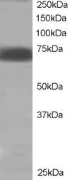
ARG63447 anti-SHP2 antibody WB image
Western blot: Human muscle lysate (RIPA buffer, 35 µg total protein per lane) stained with ARG63447 anti-SHP2 antibody at 2 µg/ml dilution.
-
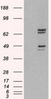
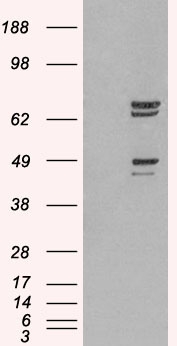
ARG63447 anti-SHP2 antibody WB image
Western blot: 1). Mock transfection; 2) PTPN11 (RC220029) expressing plasmid transfected HEK293 cell lysate standed with ARG63447 anti-SHP2 antibody.
-

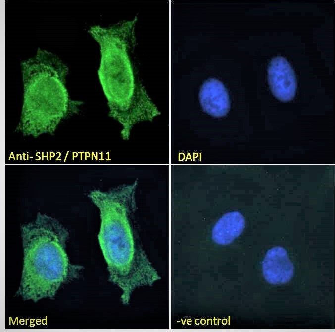
ARG63447 anti-SHP2 antibody ICC/IF image
Immunofluorescence: Paraformaldehyde fixed HeLa cells permeabilized with 0.15% Triton. Cells were stained with ARG63447 anti-SHP2 antibody (green) at 10 µg/ml dilution for 1 hour. DAPI (blue) for nuclear staining. Negative control: Unimmunized goat IgG (green) at 10 µg/ml dilution.
-
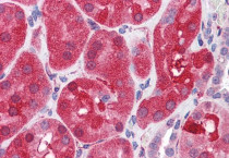
ARG63447 anti-SHP2 antibody IHC-P image
Immunohistochemistry: Paraffin-embedded Human kidney tissue. Antigen Retrieval: Steam tissue section in Citrate buffer (pH 6.0). The tissue section was stained with ARG63447 anti-SHP2 antibody at 3.75 µg/ml dilution followed by AP-staining.
-

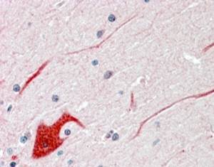
ARG63447 anti-SHP2 antibody IHC-P image
Immunohistochemistry: Paraffin embedded Human Cerebellum (Steamed antigen retrieval with citrate buffer pH 6) stained with ARG63447 anti-SHP2 antibody at 3.8 µg/ml dilution followed by AP-staining.
-

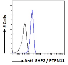
ARG63447 anti-SHP2 antibody FACS image
Flow Cytometry: Paraformaldehyde-fixed A431 cells permeabilized with 0.5% Triton. Cells were stained with ARG63447 anti-SHP2 antibody (blue line) at 10 µg/ml dilution for 1 hour, followed by incubation with Alexa FluorR 488 labelled secondary antibody. IgG control: Unimmunized goat IgG (black line).