ARG10057
anti-SHP1 antibody [PTY11]
anti-SHP1 antibody [PTY11] for ELISA,ICC/IF,Western blot and Human,Mouse
Cancer antibody; Cell Biology and Cellular Response antibody; Developmental Biology antibody; Signaling Transduction antibody

Overview
| Product Description | Mouse Monoclonal antibody [PTY11] recognizes SHP1 |
|---|---|
| Tested Reactivity | Hu, Ms |
| Tested Application | ELISA, ICC/IF, WB |
| Host | Mouse |
| Clonality | Monoclonal |
| Clone | PTY11 |
| Isotype | IgG1, kappa |
| Target Name | SHP1 |
| Antigen Species | Human |
| Immunogen | Purified recombinant protein corresponding to full-length PTP1-C |
| Conjugation | Un-conjugated |
| Alternate Names | HCP; Hematopoietic cell protein-tyrosine phosphatase; Protein-tyrosine phosphatase SHP-1; SH-PTP1; PTP-1C; HPTP1C; HCPH; Tyrosine-protein phosphatase non-receptor type 6; SHP-1; EC 3.1.3.48; SHP1; SHP-1L; Protein-tyrosine phosphatase 1C |
Application Instructions
| Application Note | * The dilutions indicate recommended starting dilutions and the optimal dilutions or concentrations should be determined by the scientist. |
|---|---|
| Positive Control | add mouse based on RD tested data, CCW, 2017-11-14; |
Properties
| Form | Liquid |
|---|---|
| Purification | Protein G affinity purified |
| Buffer | 0.01M PBS (pH 7.2) |
| Concentration | 1 mg/ml |
| Storage Instruction | For continuous use, store undiluted antibody at 2-8°C for up to a week. For long-term storage, aliquot and store at -20°C or below. Storage in frost free freezers is not recommended. Avoid repeated freeze/thaw cycles. Suggest spin the vial prior to opening. The antibody solution should be gently mixed before use. |
| Note | For laboratory research only, not for drug, diagnostic or other use. |
Bioinformation
| Database Links |
Swiss-port # P29350 Human Tyrosine-protein phosphatase non-receptor type 6 Swiss-port # P29351 Mouse Tyrosine-protein phosphatase non-receptor type 6 |
|---|---|
| Gene Symbol | PTPN6 |
| Gene Full Name | protein tyrosine phosphatase, non-receptor type 6 |
| Background | The protein encoded by this gene is a member of the protein tyrosine phosphatase (PTP) family. PTPs are known to be signaling molecules that regulate a variety of cellular processes including cell growth, differentiation, mitotic cycle, and oncogenic transformation. N-terminal part of this PTP contains two tandem Src homolog (SH2) domains, which act as protein phospho-tyrosine binding domains, and mediate the interaction of this PTP with its substrates. This PTP is expressed primarily in hematopoietic cells, and functions as an important regulator of multiple signaling pathways in hematopoietic cells. This PTP has been shown to interact with, and dephosphorylate a wide spectrum of phospho-proteins involved in hematopoietic cell signaling. Multiple alternatively spliced variants of this gene, which encode distinct isoforms, have been reported. [provided by RefSeq, Jul 2008] |
| Function | Modulates signaling by tyrosine phosphorylated cell surface receptors such as KIT and the EGF receptor/EGFR. The SH2 regions may interact with other cellular components to modulate its own phosphatase activity against interacting substrates. Together with MTUS1, induces UBE2V2 expression upon angiotensin II stimulation. Plays a key role in hematopoiesis. [UniProt] |
| Highlight | Related products: SHP1 antibodies; Anti-Mouse IgG secondary antibodies; Related news: Tools for studying H. pylori diseases |
| Research Area | Cancer antibody; Cell Biology and Cellular Response antibody; Developmental Biology antibody; Signaling Transduction antibody |
| Calculated MW | 68 kDa |
| PTM | Phosphorylated on tyrosine residues. Binding of KITLG/SCF to KIT increases tyrosine phosphorylation (By similarity). Phosphorylation at Tyr-564 enhances phosphatase activity. |
Images (1) Click the Picture to Zoom In
-
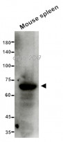
ARG10057 anti-SHP1 antibody [PTY11] WB image
Western blot: 15 µg of Mouse spleen lysate stained with ARG10057 anti-SHP1 antibody [PTY11] at 1:500 dilution.

Customer's Feedback
Good

Review for anti-SHP1 antibody [PTY11]
Application:WB
Sample:Mouse spleen
Sample Loading Amount:15 µg
Primary Antibody Dilution Factor:1:500
Primary Antibody Incubation Time:overnight
Primary Antibody Incubation Temperature:4 ºC

Clone References
Angiotensin II and endothelin-1 augment the vascular complications of diabetes via JAK2 activation.
WB / Rat


Defective hepatic response to interferon and activation of suppressor of cytokine signaling 3 in chronic hepatitis C.


