ARG64175
anti-SHP1 antibody
anti-SHP1 antibody for IHC-Formalin-fixed paraffin-embedded sections,Western blot and Human,Mouse,Rat
Cancer antibody; Cell Biology and Cellular Response antibody; Developmental Biology antibody; Signaling Transduction antibody
Overview
| Product Description | Goat Polyclonal antibody recognizes SHP1 |
|---|---|
| Tested Reactivity | Hu, Ms, Rat |
| Predict Reactivity | Dog |
| Tested Application | IHC-P, WB |
| Specificity | This antibody is expected to recognise both reported isoforms (NP_536858.1and NP_002822.2). _x000D_Please note this antibody was designed using the mouse sequence, which differs by 1 amino acid from the human sequence. |
| Host | Goat |
| Clonality | Polyclonal |
| Isotype | IgG |
| Target Name | SHP1 |
| Antigen Species | Human |
| Immunogen | C-KASRTSSKHKEE |
| Conjugation | Un-conjugated |
| Alternate Names | HCP; Hematopoietic cell protein-tyrosine phosphatase; Protein-tyrosine phosphatase SHP-1; SH-PTP1; PTP-1C; HPTP1C; HCPH; Tyrosine-protein phosphatase non-receptor type 6; SHP-1; EC 3.1.3.48; SHP1; SHP-1L; Protein-tyrosine phosphatase 1C |
Application Instructions
| Application Suggestion |
|
||||||
|---|---|---|---|---|---|---|---|
| Application Note | WB: Recommend incubate at RT for 1h. IHC-P: Antigen Retrieval: Steam tissue section in Citrate buffer (pH 6.0). * The dilutions indicate recommended starting dilutions and the optimal dilutions or concentrations should be determined by the scientist. |
Properties
| Form | Liquid |
|---|---|
| Purification | Purified from goat serum by antigen affinity chromatography. |
| Buffer | Tris saline (pH 7.3), 0.02% Sodium azide and 0.5% BSA. |
| Preservative | 0.02% Sodium azide |
| Stabilizer | 0.5% BSA |
| Concentration | 0.5 mg/ml |
| Storage Instruction | For continuous use, store undiluted antibody at 2-8°C for up to a week. For long-term storage, aliquot and store at -20°C or below. Storage in frost free freezers is not recommended. Avoid repeated freeze/thaw cycles. Suggest spin the vial prior to opening. The antibody solution should be gently mixed before use. |
| Note | For laboratory research only, not for drug, diagnostic or other use. |
Bioinformation
| Database Links | |
|---|---|
| Background | The protein encoded by this gene is a member of the protein tyrosine phosphatase (PTP) family. PTPs are known to be signaling molecules that regulate a variety of cellular processes including cell growth, differentiation, mitotic cycle, and oncogenic transformation. N-terminal part of this PTP contains two tandem Src homolog (SH2) domains, which act as protein phospho-tyrosine binding domains, and mediate the interaction of this PTP with its substrates. This PTP is expressed primarily in hematopoietic cells, and functions as an important regulator of multiple signaling pathways in hematopoietic cells. This PTP has been shown to interact with, and dephosphorylate a wide spectrum of phospho-proteins involved in hematopoietic cell signaling. Multiple alternatively spliced variants of this gene, which encode distinct isoforms, have been reported. [provided by RefSeq, Jul 2008] |
| Research Area | Cancer antibody; Cell Biology and Cellular Response antibody; Developmental Biology antibody; Signaling Transduction antibody |
| Calculated MW | 68 kDa |
| PTM | Phosphorylated on tyrosine residues. Binding of KITLG/SCF to KIT increases tyrosine phosphorylation (By similarity). Phosphorylation at Tyr-564 enhances phosphatase activity. |
Images (3) Click the Picture to Zoom In
-
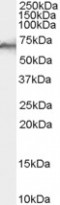
ARG64175 anti-SHP1 antibody WB image
Western blot: Human Liver lysate (35 µg protein in RIPA buffer) stained with ARG64175 anti-SHP1 antibody at 0.1 µg/ml dilution.
-

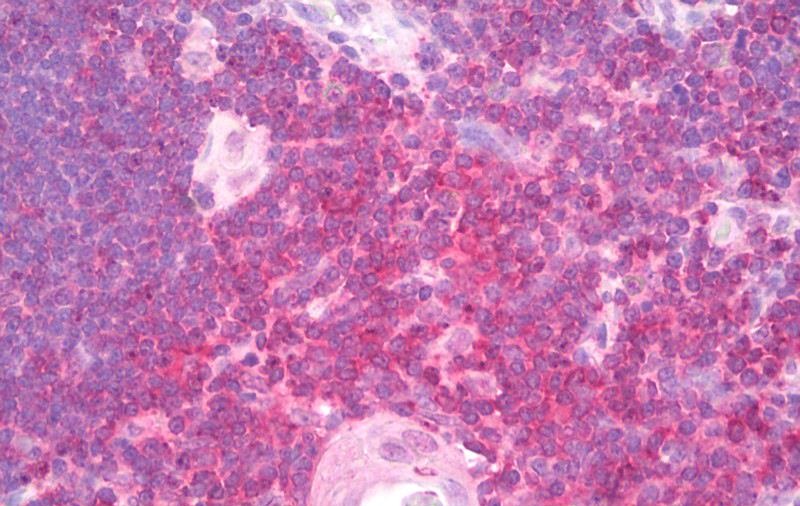
ARG64175 anti-SHP1 antibody IHC-P image
Immunohistochemistry: Paraffin-embedded Human thyroid tissue. Antigen Retrieval: Steam tissue section in Citrate buffer (pH 6.0). The tissue section was stained with ARG64175 anti-SHP1 antibody at 5 µg/ml dilution followed by AP-staining.
-
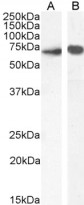
ARG64175 anti-SHP1 antibody WB image
Western blot: 35 µg of Mouse thymus (A) and Rat thymus (B) lysates (in RIPA buffer) stained with ARG64175 anti-SHP1 antibody at 0.01 µg/ml (A) and 0.1 µg/ml (B) dilutions and incubated at RT for 1 hour.