ARG55768
anti-SHB antibody
anti-SHB antibody for Flow cytometry,IHC-Formalin-fixed paraffin-embedded sections,Western blot and Human
Overview
| Product Description | Rabbit Polyclonal antibody recognizes SHB |
|---|---|
| Tested Reactivity | Hu |
| Tested Application | FACS, IHC-P, WB |
| Host | Rabbit |
| Clonality | Polyclonal |
| Isotype | IgG |
| Target Name | SHB |
| Antigen Species | Human |
| Immunogen | KLH-conjugated synthetic peptide corresponding to aa. 250-290 of Human SHB. |
| Conjugation | Un-conjugated |
| Alternate Names | SH2 domain-containing adapter protein B; bA3J10.2 |
Application Instructions
| Application Suggestion |
|
||||||||
|---|---|---|---|---|---|---|---|---|---|
| Application Note | * The dilutions indicate recommended starting dilutions and the optimal dilutions or concentrations should be determined by the scientist. | ||||||||
| Positive Control | HepG2 |
Properties
| Form | Liquid |
|---|---|
| Purification | Purification with Protein A and immunogen peptide. |
| Buffer | PBS and 0.09% (W/V) sodium azide. |
| Preservative | 0.09% (W/V) sodium azide. |
| Storage Instruction | For continuous use, store undiluted antibody at 2-8°C for up to a week. For long-term storage, aliquot and store at -20°C or below. Storage in frost free freezers is not recommended. Avoid repeated freeze/thaw cycles. Suggest spin the vial prior to opening. The antibody solution should be gently mixed before use. |
| Note | For laboratory research only, not for drug, diagnostic or other use. |
Bioinformation
| Database Links |
Swiss-port # Q15464 Human SH2 domain-containing adapter protein B |
|---|---|
| Gene Symbol | SHB |
| Gene Full Name | Src homology 2 domain containing adaptor protein B |
| Function | Adapter protein which regulates several signal transduction cascades by linking activated receptors to downstream signaling components. May play a role in angiogenesis by regulating FGFR1, VEGFR2 and PDGFR signaling. May also play a role in T-cell antigen receptor/TCR signaling, interleukin-2 signaling, apoptosis and neuronal cells differentiation by mediating basic-FGF and NGF-induced signaling cascades. May also regulate IRS1 and IRS2 signaling in insulin-producing cells. [UniProt] |
| Cellular Localization | Cytoplasm. Cell membrane; Peripheral membrane protein; Cytoplasmic side. Note=Associates with membrane lipid rafts upon TCR stimulation |
| Calculated MW | 55 kDa |
| PTM | Phosphorylated upon PDGFRA, PDGFRB, TCR, IL2 receptor, FGFR1 or VEGFR2 activation. |
Images (3) Click the Picture to Zoom In
-
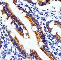
ARG55768 anti-SHB antibody IHC-P image
Immunohistochemistry: Paraffin-embedded Human small intestine tissue stained with ARG55768 anti-SHB antibody at 1:25 dilution.
-

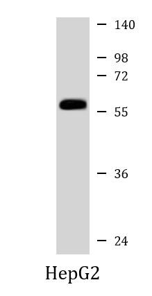
ARG55768 anti-SHB antibody WB image
Western blot: 35 µg of HepG2 cell lysate stained with ARG55768 anti-SHB antibody at 1:1000 dilution.
-
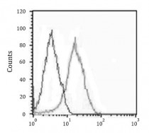
ARG55768 anti-SHB antibody FACS image
Flow Cytometry: HepG2 cells stained with ARG55768 anti-SHB antibody (right histogram) at 1:25 dilution or isotype control antibody (left histogram), followed by incubation with Alexa Fluor® 488 labelled secondary antibody.