ARG40998
anti-SDHB antibody [2I3]
anti-SDHB antibody [2I3] for Flow cytometry,IHC-Formalin-fixed paraffin-embedded sections,Western blot and Human,Mouse,Rat
Overview
| Product Description | Mouse Monoclonal antibody [2I3] recognizes SDHB |
|---|---|
| Tested Reactivity | Hu, Ms, Rat |
| Tested Application | FACS, IHC-P, WB |
| Host | Mouse |
| Clonality | Monoclonal |
| Clone | 2I3 |
| Isotype | IgG1 |
| Target Name | SDHB |
| Antigen Species | Human |
| Immunogen | Recombinant protein corresponding to A29-V280 of Human SDHB. |
| Conjugation | Un-conjugated |
| Alternate Names | Ip; SDH2; SDH1; IP; EC 1.3.5.1; PGL4; CWS2; Succinate dehydrogenase [ubiquinone] iron-sulfur subunit, mitochondrial; SDHIP; Iron-sulfur subunit of complex II; SDH |
Application Instructions
| Application Suggestion |
|
||||||||
|---|---|---|---|---|---|---|---|---|---|
| Application Note | IHC-P: Antigen Retrieval: Heat mediation was performed in Citrate buffer (pH 6.0, epitope retrieval solution) for 20 min. * The dilutions indicate recommended starting dilutions and the optimal dilutions or concentrations should be determined by the scientist. |
||||||||
| Observed Size | 29 kDa |
Properties
| Form | Liquid |
|---|---|
| Purification | Affinity purification with immunogen. |
| Buffer | 0.2% Na2HPO4, 0.9% NaCl, 0.05% Sodium azide and 4% Trehalose. |
| Preservative | 0.05% Sodium azide |
| Stabilizer | 4% Trehalose |
| Concentration | 0.5 mg/ml |
| Storage Instruction | For continuous use, store undiluted antibody at 2-8°C for up to a week. For long-term storage, aliquot and store at -20°C or below. Storage in frost free freezers is not recommended. Avoid repeated freeze/thaw cycles. Suggest spin the vial prior to opening. The antibody solution should be gently mixed before use. |
| Note | For laboratory research only, not for drug, diagnostic or other use. |
Bioinformation
| Database Links | |
|---|---|
| Gene Symbol | SDHB |
| Gene Full Name | succinate dehydrogenase complex, subunit B, iron sulfur (Ip) |
| Background | Complex II of the respiratory chain, which is specifically involved in the oxidation of succinate, carries electrons from FADH to CoQ. The complex is composed of four nuclear-encoded subunits and is localized in the mitochondrial inner membrane. The iron-sulfur subunit is highly conserved and contains three cysteine-rich clusters which may comprise the iron-sulfur centers of the enzyme. Sporadic and familial mutations in this gene result in paragangliomas and pheochromocytoma, and support a link between mitochondrial dysfunction and tumorigenesis. [provided by RefSeq, Jul 2008] |
| Function | Iron-sulfur protein (IP) subunit of succinate dehydrogenase (SDH) that is involved in complex II of the mitochondrial electron transport chain and is responsible for transferring electrons from succinate to ubiquinone (coenzyme Q). [UniProt] |
| Cellular Localization | Mitochondrion inner membrane; Peripheral membrane protein; Matrix side. [UniProt] |
| Calculated MW | 32 kDa |
Images (7) Click the Picture to Zoom In
-
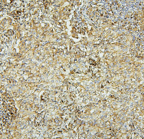
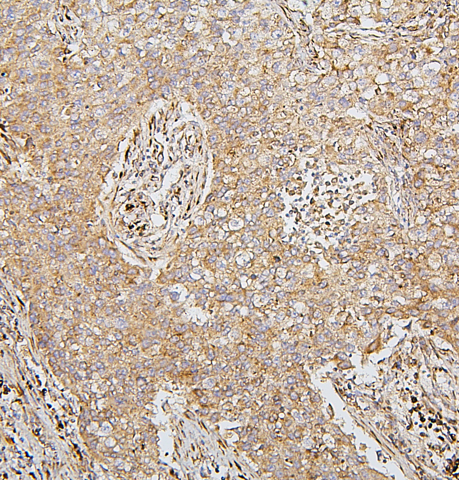
ARG40998 anti-SDHB antibody [2I3] IHC-P image
Immunohistochemistry: Paraffin-embedded Human lung cancer tissue. Antigen Retrieval: Heat mediation was performed in Citrate buffer (pH 6.0, epitope retrieval solution) for 20 min. The tissue section was blocked with 10% goat serum. The tissue section was then stained with ARG40998 anti-SDHB antibody [2I3] at 1 µg/ml dilution, overnight at 4°C.
-
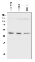
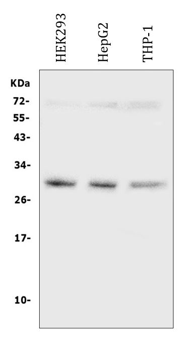
ARG40998 anti-SDHB antibody [2I3] WB image
Western blot: 50 µg of samples under reducing conditions. HEK293, HepG2 and THP-1 whole cell lysates stained with ARG40998 anti-SDHB antibody [2I3] at 0.5 µg/ml, overnight at 4°C.
-
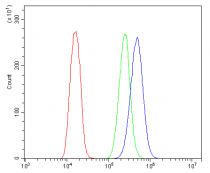
ARG40998 anti-SDHB antibody [2I3] FACS image
Flow Cytometry: U-87MG cells were blocked with 10% normal goat serum and then stained with ARG40998 anti-SDHB antibody [2I3] (blue) at 1 µg/10^6 cells for 30 min at 20°C, followed by incubation with DyLight®488 labelled secondary antibody. Isotype control antibody (green) was Mouse IgG (1 µg/10^6 cells) used under the same conditions. Unlabelled sample (red) was also used as a control.
-

ARG40998 anti-SDHB antibody [2I3] IHC-P image
Immunohistochemistry: Paraffin-embedded Human lung cancer tissue. Antigen Retrieval: Heat mediation was performed in Citrate buffer (pH 6.0, epitope retrieval solution) for 20 min. The tissue section was blocked with 10% goat serum. The tissue section was then stained with ARG40998 anti-SDHB antibody [2I3] at 1 µg/ml dilution, overnight at 4°C.
-
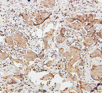
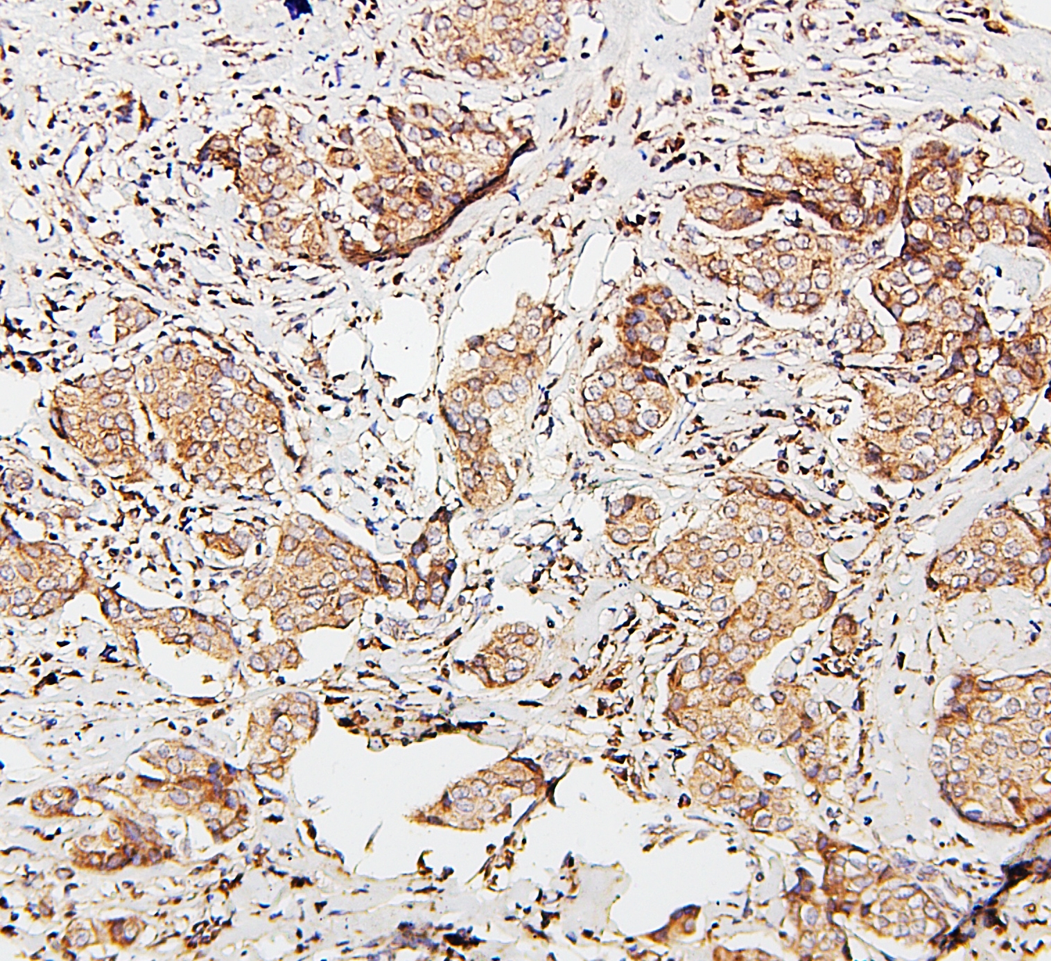
ARG40998 anti-SDHB antibody [2I3] IHC-P image
Immunohistochemistry: Paraffin-embedded Human mammary cancer tissue. Antigen Retrieval: Heat mediation was performed in Citrate buffer (pH 6.0, epitope retrieval solution) for 20 min. The tissue section was blocked with 10% goat serum. The tissue section was then stained with ARG40998 anti-SDHB antibody [2I3] at 1 µg/ml dilution, overnight at 4°C.
-
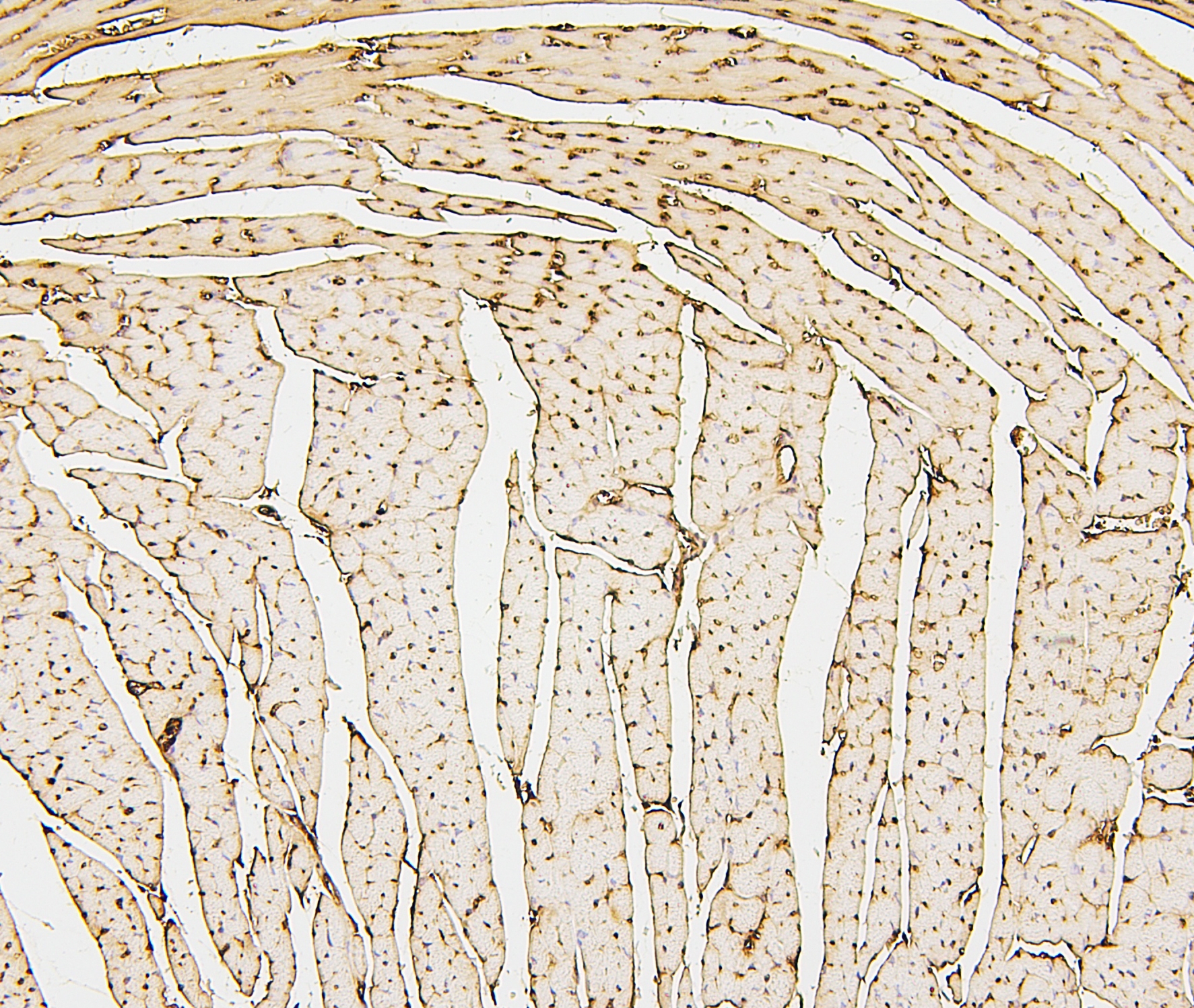
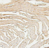
ARG40998 anti-SDHB antibody [2I3] IHC-P image
Immunohistochemistry: Paraffin-embedded Mouse cardiac muscle tissue. Antigen Retrieval: Heat mediation was performed in Citrate buffer (pH 6.0, epitope retrieval solution) for 20 min. The tissue section was blocked with 10% goat serum. The tissue section was then stained with ARG40998 anti-SDHB antibody [2I3] at 1 µg/ml dilution, overnight at 4°C.
-
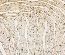
ARG40998 anti-SDHB antibody [2I3] IHC-P image
Immunohistochemistry: Paraffin-embedded Rat cardiac muscle tissue. Antigen Retrieval: Heat mediation was performed in Citrate buffer (pH 6.0, epitope retrieval solution) for 20 min. The tissue section was blocked with 10% goat serum. The tissue section was then stained with ARG40998 anti-SDHB antibody [2I3] at 1 µg/ml dilution, overnight at 4°C.