ARG43282
anti-SARS-CoV-2 Spike protein (S2) antibody
anti-SARS-CoV-2 Spike protein (S2) antibody for IHC-Formalin-fixed paraffin-embedded sections,Western blot and Virus
Overview
| Product Description | Rabbit Polyclonal antibody recognizes SARS-CoV-2 Spike protein (S2) |
|---|---|
| Tested Reactivity | Virus |
| Tested Application | IHC-P, WB |
| Host | Rabbit |
| Clonality | Polyclonal |
| Isotype | IgG |
| Target Name | SARS-CoV-2 Spike protein (S2) |
| Antigen Species | Virus |
| Immunogen | A 16-amino acid synthetic peptide within aa. 1130-1180 of SARS-CoV-2 Spike protein. |
| Conjugation | Un-conjugated |
Application Instructions
| Application Suggestion |
|
||||||
|---|---|---|---|---|---|---|---|
| Application Note | IHC-P: Antigen Retrieval: Heat mediation was performed in Citrate buffer (pH 6.0). * The dilutions indicate recommended starting dilutions and the optimal dilutions or concentrations should be determined by the scientist. |
Properties
| Form | Liquid |
|---|---|
| Purification | Affinity purification with immunogen. |
| Buffer | PBS and 0.02% Sodium azide. |
| Preservative | 0.02% Sodium azide |
| Concentration | 1 mg/ml |
| Storage Instruction | For continuous use, store undiluted antibody at 2-8°C for up to a week. For long-term storage, aliquot and store at -20°C or below. Storage in frost free freezers is not recommended. Avoid repeated freeze/thaw cycles. Suggest spin the vial prior to opening. The antibody solution should be gently mixed before use. |
| Note | For laboratory research only, not for drug, diagnostic or other use. |
Bioinformation
| Background | Severe acute respiratory syndrome coronavirus 2 (SARS-CoV-2) is an enveloped, positive-sense, single-stranded RNA virus that causes coronavirus disease 2019 (COVID-19). Virus particles include the RNA genetic material and structural proteins needed for invasion of host cells. Once inside the cell the infecting RNA is used to encode structural proteins that make up virus particles, nonstructural proteins that direct virus assembly, transcription, replication and host control and accessory proteins whose function has not been determined.~ The structural proteins of SARS-CoV-2 include the envelope protein (E), spike or surface glycoprotein (S), membrane protein (M) and the nucleocapsid protein (N). The spike glycoprotein is found on the outside of the virus particle and gives coronavirus viruses their crown-like appearance. This glycoprotein mediates attachment of the virus particle and entry into the host cell. S protein is an important target for vaccine development, antibody therapies and diagnostic antigen-based tests. |
|---|---|
| Function | mediates fusion of the virion and cellular membranes by acting as a class I viral fusion protein. Under the current model, the protein has at least three conformational states: pre-fusion native state, pre-hairpin intermediate state, and post-fusion hairpin state. During viral and target cell membrane fusion, the coiled coil regions (heptad repeats) assume a trimer-of-hairpins structure, positioning the fusion peptide in close proximity to the C-terminal region of the ectodomain. The formation of this structure appears to drive apposition and subsequent fusion of viral and target cell membranes. [UniProt] |
Images (3) Click the Picture to Zoom In
-
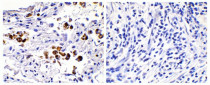
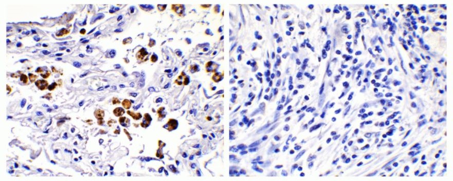
ARG43282 anti-SARS-CoV-2 Spike protein (S2) antibody IHC-P image
Immunohistochemistry: Paraffin-embedded COVID-19 patient lung tissue. Tissue was fixed with formaldehyde and blocked with 10% serum for 1 hour at RT. Antigen Retrieval: Heat mediation was performed in Citrate buffer (pH 6.0). The tissue section was stained with ARG43282 anti-SARS-CoV-2 Spike protein (S2) antibody at 0.1 µg/ml dilution, overnight at 4°C.
Strong signal of SARS-COV-2 spike protein was observed in macrophage of COVID-19 patient lung (left), but not in non-COVID-19 patient lung (right).
-

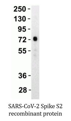
ARG43282 anti-SARS-CoV-2 Spike protein (S2) antibody WB image
Western blot: 30 ng of SARS-CoV-2 Spike S2 recombinant protein stained with ARG43282 anti-SARS-CoV-2 Spike protein (S2) antibody at 1 µg/ml dilution, 1 hour incubation at RT in 5% NFDM/TBST.
-
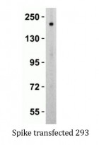
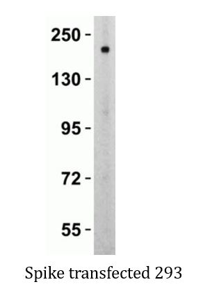
ARG43282 anti-SARS-CoV-2 Spike protein (S2) antibody WB image
Western blot: Spike transfected 293 cells. 10 µg of cell lysate stained with ARG43282 anti-SARS-CoV-2 Spike protein (S2) antibody at 1 µg/ml dilution, 1 hour incubation at RT in 5% NFDM/TBST.