ARG55752
anti-S6 Ribosomal Protein antibody
anti-S6 Ribosomal Protein antibody for Flow cytometry,ICC/IF,IHC-Formalin-fixed paraffin-embedded sections,Western blot and Human,Mouse,Rat
Overview
| Product Description | Mouse Monoclonal antibody recognizes S6 Ribosomal Protein |
|---|---|
| Tested Reactivity | Hu, Ms, Rat |
| Tested Application | FACS, ICC/IF, IHC-P, WB |
| Host | Mouse |
| Clonality | Monoclonal |
| Clone | 981CT2.1.1 |
| Isotype | IgG1 |
| Target Name | S6 Ribosomal Protein |
| Antigen Species | Human |
| Immunogen | Human S6 Ribosomal recombinant protein |
| Conjugation | Un-conjugated |
| Alternate Names | Phosphoprotein NP33; 40S ribosomal protein S6; S6 |
Application Instructions
| Application Suggestion |
|
||||||||||
|---|---|---|---|---|---|---|---|---|---|---|---|
| Application Note | * The dilutions indicate recommended starting dilutions and the optimal dilutions or concentrations should be determined by the scientist. | ||||||||||
| Positive Control | 293 |
Properties
| Form | Liquid |
|---|---|
| Purification | Purification with Protein G. |
| Buffer | PBS and 0.09% (W/V) Sodium azide. |
| Preservative | 0.09% (W/V) Sodium azide. |
| Storage Instruction | For continuous use, store undiluted antibody at 2-8°C for up to a week. For long-term storage, aliquot and store at -20°C or below. Storage in frost free freezers is not recommended. Avoid repeated freeze/thaw cycles. Suggest spin the vial prior to opening. The antibody solution should be gently mixed before use. |
| Note | For laboratory research only, not for drug, diagnostic or other use. |
Bioinformation
| Database Links | |
|---|---|
| Gene Symbol | RPS6 |
| Gene Full Name | ribosomal protein S6 |
| Background | Ribosomes, the organelles that catalyze protein synthesis, consist of a small 40S subunit and a large 60S subunit. Together these subunits are composed of 4 RNA species and approximately 80 structurally distinct proteins. This gene encodes a cytoplasmic ribosomal protein that is a component of the 40S subunit. The protein belongs to the S6E family of ribosomal proteins. It is the major substrate of protein kinases in the ribosome, with subsets of five C-terminal serine residues phosphorylated by different protein kinases. Phosphorylation is induced by a wide range of stimuli, including growth factors, tumor-promoting agents, and mitogens. Dephosphorylation occurs at growth arrest. The protein may contribute to the control of cell growth and proliferation through the selective translation of particular classes of mRNA. As is typical for genes encoding ribosomal proteins, there are multiple processed pseudogenes of this gene dispersed through the genome. [provided by RefSeq, Jul 2008] |
| Function | May play an important role in controlling cell growth and proliferation through the selective translation of particular classes of mRNA. [UniProt] |
| Calculated MW | 29 kDa |
| PTM | Ribosomal protein S6 is the major substrate of protein kinases in eukaryote ribosomes. The phosphorylation is stimulated by growth factors, tumor promoting agents, and mitogens. It is dephosphorylated at growth arrest. Phosphorylated at Ser-235 and Ser-236 by RPS6KA1 and RPS6KA3; phosphorylation at these sites facilitates the assembly of the preinitiation complex. |
Images (4) Click the Picture to Zoom In
-
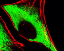
ARG55752 anti-S6 Ribosomal Protein antibody ICC/IF image
Immunofluorescence: HeLa cells stained with ARG55752 anti-S6 Ribosomal Protein antibody (green) at 1:25 dilution. Cytoplasmic actin was counterstained with Alexa Fluor® 555 conjugated with Phalloidin (red).
-

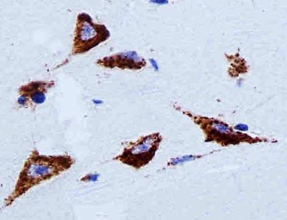
ARG55752 anti-S6 Ribosomal Protein antibody IHC-P image
Immunohistochemistry: Paraffin-embedded Human brain tissue stained with ARG55752 anti-S6 Ribosomal Protein antibody at 1:25 dilution.
-

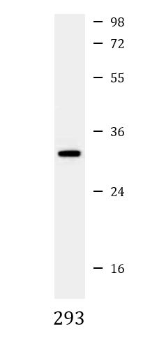
ARG55752 anti-S6 Ribosomal Protein antibody WB image
Western blot: 35 µg of 293 cell lysate stained with ARG55752 anti-S6 Ribosomal Protein antibody at 1:2000 dilution.
-

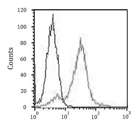
ARG55752 anti-S6 Ribosomal Protein antibody FACS image
Flow Cytometry: HeLa cells stained with ARG55752 anti-S6 Ribosomal Protein antibody (right histogram) at 1:25 dilution or isotype control antibody (left histogram), followed by incubation with Alexa Fluor® 488 labelled secondary antibody.