ARG64813
anti-S100A9 antibody
anti-S100A9 antibody for Flow cytometry,ICC/IF,IHC-Formalin-fixed paraffin-embedded sections,Western blot and Human
Cancer antibody; Immune System antibody; Signaling Transduction antibody
Overview
| Product Description | Goat Polyclonal antibody recognizes S100A9 |
|---|---|
| Tested Reactivity | Hu |
| Tested Application | FACS, ICC/IF, IHC-P, WB |
| Host | Goat |
| Clonality | Polyclonal |
| Isotype | IgG |
| Target Name | S100A9 |
| Antigen Species | Human |
| Immunogen | C-DTNADKQLSFEEF |
| Conjugation | Un-conjugated |
| Alternate Names | Calgranulin-B; MRP-14; MRP14; 60B8AG; CFAG; MAC387; Calprotectin L1H subunit; NIF; MIF; p14; LIAG; Protein S100-A9; CGLB; Migration inhibitory factor-related protein 14; L1AG; Leukocyte L1 complex heavy chain; P14; CAGB; S100 calcium-binding protein A9 |
Application Instructions
| Application Suggestion |
|
||||||||||
|---|---|---|---|---|---|---|---|---|---|---|---|
| Application Note | WB: Recommend incubate at RT for 1h. IHC-P: Antigen Retrieval: Steam tissue section in Citrate buffer (pH 6.0). * The dilutions indicate recommended starting dilutions and the optimal dilutions or concentrations should be determined by the scientist. |
Properties
| Form | Liquid |
|---|---|
| Purification | Purified from goat serum by antigen affinity chromatography. |
| Buffer | Tris saline (pH 7.3), 0.02% Sodium azide and 0.5% BSA. |
| Preservative | 0.02% Sodium azide |
| Stabilizer | 0.5% BSA |
| Concentration | 0.5 mg/ml |
| Storage Instruction | For continuous use, store undiluted antibody at 2-8°C for up to a week. For long-term storage, aliquot and store at -20°C or below. Storage in frost free freezers is not recommended. Avoid repeated freeze/thaw cycles. Suggest spin the vial prior to opening. The antibody solution should be gently mixed before use. |
| Note | For laboratory research only, not for drug, diagnostic or other use. |
Bioinformation
| Database Links | |
|---|---|
| Background | The protein encoded by this gene is a member of the S100 family of proteins containing 2 EF-hand calcium-binding motifs. S100 proteins are localized in the cytoplasm and/or nucleus of a wide range of cells, and involved in the regulation of a number of cellular processes such as cell cycle progression and differentiation. S100 genes include at least 13 members which are located as a cluster on chromosome 1q21. This protein may function in the inhibition of casein kinase and altered expression of this protein is associated with the disease cystic fibrosis. [provided by RefSeq, Jul 2008] |
| Highlight | Related products: S100A antibodies; S100A ELISA Kits; Anti-Goat IgG secondary antibodies; Related news: HMGB1, a biomarker and therapeutic target in COVID-19 |
| Research Area | Cancer antibody; Immune System antibody; Signaling Transduction antibody |
| Calculated MW | 13 kDa |
| PTM | Phosphorylated. Phosphorylation inhibits activation of tubulin polymerization. S-nitrosylation of Cys-3 is implicated in LDL(ox)-induced S-nitrosylation of GAPDH at 'Cys-247' through a transnitrosylase mechanism involving a iNOS-S100A8/9 complex (PubMed:25417112). |
Images (6) Click the Picture to Zoom In
-
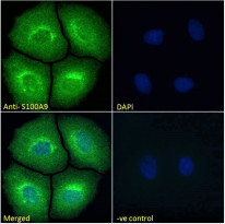
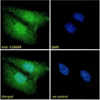
ARG64813 anti-S100A9 antibody ICC/IF image
Immunofluorescence: Paraformaldehyde-fixed MCF7 cells permeabilized with 0.15% Triton. Cells were stained with ARG64813 anti-S100A9 antibody (green) at 10 µg/ml dilution for 1 hour. DAPI (blue) for nuclear staining. Negative control: Unimmunized Goat IgG (green) at 10 µg/ml dilution.
-
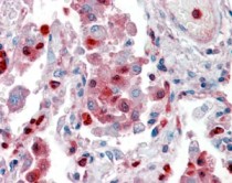
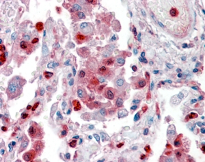
ARG64813 anti-S100A9 antibody IHC-P image
Immunohistochemistry: Paraffin-embedded Human lung tissue. Antigen Retrieval: Steam tissue section in Citrate buffer (pH 6.0). The tissue section was stained with ARG64813 anti-S100A9 antibody at 2.5 µg/ml dilution followed by AP-staining.
-

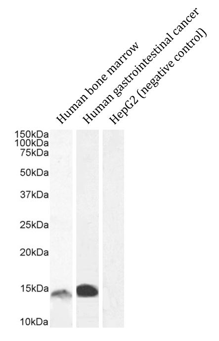
ARG64813 anti-S100A9 antibody WB image
Western blot: 35 µg of Human bone marrow, Human gastrointestinal cancer and HepG2 cell lysates (negative control) stained with ARG64813 anti-S100A9 antibody at 1 µg/ml (bone marrow) and 0.5 µg/ml (gastrointestinal cancer) dilutions and incubated at RT for 1 hour.
-
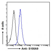
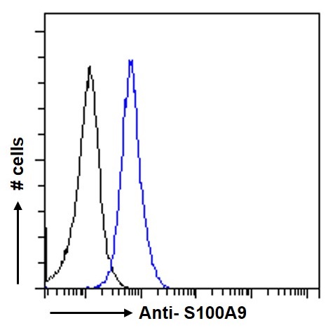
ARG64813 anti-S100A9 antibody FACS image
Flow Cytometry: Paraformaldehyde-fixed MCF7 cells permeabilized with 0.5% Triton. Cells were stained with ARG64813 anti-S100A9 antibody (blue line) at 10 µg/ml dilution for 1 hour, followed by incubation with Alexa Fluor® 488 labelled secondary antibody. IgG control: Unimmunized Goat IgG (black line), followed by incubation with Alexa Fluor® 488 labelled secondary antibody.
-
ARG64813 anti-S100A9 antibody ICC/IF image
Immunofluorescence: Paraformaldehyde-fixed U2OS cells permeabilized with 0.15% Triton. Cells were stained with ARG64813 anti-S100A9 antibody (green) at 10 µg/ml dilution for 1 hour. DAPI (blue) for nuclear staining. Negative control: Unimmunized Goat IgG (green) at 10 µg/ml dilution.
-
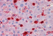
ARG64813 anti-S100A9 antibody IHC-P image
Immunohistochemistry: Paraffin-embedded Human liver tissue. Antigen Retrieval: Steam tissue section in Citrate buffer (pH 6.0). The tissue section was stained with ARG64813 anti-S100A9 antibody at 2.5 µg/ml dilution followed by AP-staining.