ARG11145
anti-Rhodopsin antibody [B630]
anti-Rhodopsin antibody [B630] for ICC/IF,IHC-Frozen sections,Western blot and Human,Mouse,Rat,Cow,Horse,Pig
Overview
| Product Description | Mouse Monoclonal antibody [B630] recognizes Rhodopsin |
|---|---|
| Tested Reactivity | Hu, Ms, Rat, Cow, Hrs, Pig |
| Tested Application | ICC/IF, IHC-Fr, WB |
| Host | Mouse |
| Clonality | Monoclonal |
| Clone | B630 |
| Isotype | IgG1 |
| Target Name | Rhodopsin |
| Antigen Species | Bovine |
| Immunogen | Purified bovine rhodopsin. |
| Conjugation | Un-conjugated |
| Alternate Names | Rhodopsin; Opsin-2; CSNBAD1; RP4; OPN2 |
Application Instructions
| Application Suggestion |
|
||||||||
|---|---|---|---|---|---|---|---|---|---|
| Application Note | * The dilutions indicate recommended starting dilutions and the optimal dilutions or concentrations should be determined by the scientist. | ||||||||
| Observed Size | ~ 33 kDa |
Properties
| Form | Liquid |
|---|---|
| Purification | Purified |
| Buffer | PBS, 5 mM Sodium azide and 50% Glycerol. |
| Preservative | 5 mM Sodium azide |
| Stabilizer | 50% Glycerol |
| Concentration | 1 mg/ml |
| Storage Instruction | For continuous use, store undiluted antibody at 2-8°C for up to a week. For long-term storage, aliquot and store at -20°C. Storage in frost free freezers is not recommended. Avoid repeated freeze/thaw cycles. Suggest spin the vial prior to opening. The antibody solution should be gently mixed before use. |
| Note | For laboratory research only, not for drug, diagnostic or other use. |
Bioinformation
| Database Links | |
|---|---|
| Gene Symbol | RHO |
| Gene Full Name | rhodopsin |
| Background | The protein encoded by this gene is found in rod cells in the back of the eye and is essential for vision in low-light conditions. The encoded protein binds to 11-cis retinal and is activated when light hits the retinal molecule. Defects in this gene are a cause of congenital stationary night blindness. [provided by RefSeq, Aug 2017] |
| Function | Photoreceptor required for image-forming vision at low light intensity (PubMed:8107847, PubMed:7846071). Required for photoreceptor cell viability after birth (PubMed:2215617, PubMed:12566452). Light-induced isomerization of the chromophore 11-cis-retinal to all-trans-retinal triggers a conformational change that activates signaling via G-proteins (PubMed:8107847, PubMed:28524165, PubMed:26200343, PubMed:28753425). Subsequent receptor phosphorylation mediates displacement of the bound G-protein alpha subunit by the arrestin SAG and terminates signaling (PubMed:28524165, PubMed:26200343). [UniProt] |
| Cellular Localization | Membrane; Multi-pass membrane protein. Cell projection, cilium, photoreceptor outer segment. Note=Synthesized in the inner segment (IS) of rod photoreceptor cells before vectorial transport to disk membranes in the rod outer segment (OS) photosensory cilia. [UniProt] |
| Calculated MW | 39 kDa |
| PTM | Phosphorylated on some or all of the serine and threonine residues present in the C-terminal region. Contains one covalently linked retinal chromophore. [UniProt] |
Images (3) Click the Picture to Zoom In
-
ARG11145 anti-Rhodopsin antibody [B630] IHC-Fr image
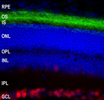
Immunohistochemistry: Frozen section of Mouse retina tissue stained with ARG11145 anti-Rhodopsin antibody [B630] (green) at 1:2000 dilution, and co-stained with ARG10712 anti-FOX3 / NeuN antibody (red) at 1:5000 dilution. Hoechst (blue) for nuclear staining.
-
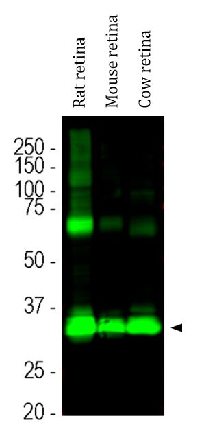
ARG11145 anti-Rhodopsin antibody [B630] WB image
Western blot: Rat retina, Mouse retina and Cow retina lysates stained with ARG11145 anti-Rhodopsin antibody [B630] at 1:5000 dilution.
The strong band at 35 kDa corresponds to rhodopsin protein. Bands at about 70 kDa and 140 kDa are presumably aggregated forms of rhodopsin.
-
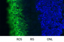
ARG11145 anti-Rhodopsin antibody [B630] IHC-Fr image
Immunohistochemistry: Frozen section of Pig retinal tissue stained with ARG11145 anti-Rhodopsin antibody [B630] (green). Hoechst (blue) for nuclear staining.
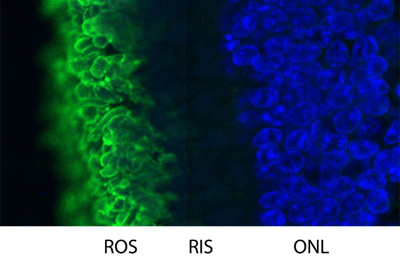
Rhodopsin is most abundant in the rod outer segments (ROS) of retina, clearly localized in rod cell membranes. The rod inner segments (RIS) and rod nuclei in the outer nuclear layer (ONL) are also seen in this image.