ARG52417
anti-Rhodopsin antibody [1D4]
anti-Rhodopsin antibody [1D4] for ELISA,ICC/IF,Immunohistochemistry (PFA perfusion fixed frozen sections),IHC-Frozen sections,IHC-Formalin-fixed paraffin-embedded sections,Immunoprecipitation,Western blot and Human,Mouse,Rat,Amphibians,Cow,Mammal,Zebrafish
Neuroscience antibody; Signaling Transduction antibody
Overview
| Product Description | Mouse Monoclonal antibody [1D4] recognizes Rhodopsin |
|---|---|
| Tested Reactivity | Hu, Ms, Rat, Amph, Cow, Mamm, Zfsh |
| Predict Reactivity | Rb |
| Tested Application | ELISA, ICC/IF, IHC-FoFr , IHC-Fr, IHC-P, IP, WB |
| Specificity | The antibody reacts to C- terminal epitope TETSQVAPA- (COOH) of rhodopsin, so it also reacts to C9 tag (TETSQVAPA). |
| Host | Mouse |
| Clonality | Monoclonal |
| Clone | 1D4 |
| Isotype | IgG1 |
| Target Name | Rhodopsin |
| Antigen Species | Bovine |
| Immunogen | Purified native bovine rhodopsin |
| Epitope | Antibody binds to the C- terminal epitope-T-E-T-S-Q-V-A-P-A- (COOH) of rhodopsin. |
| Conjugation | Un-conjugated |
| Alternate Names | Rhodopsin; Opsin-2; CSNBAD1; RP4; OPN2 |
Application Instructions
| Application Suggestion |
|
||||||||||||||||
|---|---|---|---|---|---|---|---|---|---|---|---|---|---|---|---|---|---|
| Application Note | Specific for the ~ 39k rhodopsin protein. * The dilutions indicate recommended starting dilutions and the optimal dilutions or concentrations should be determined by the scientist. |
Properties
| Form | Liquid |
|---|---|
| Purification | Protein G purified |
| Buffer | 10 mM HEPES (pH 7.5), 150 mM NaCl, 0.1 mg/ml BSA and 50% Glycerol |
| Stabilizer | 0.1 mg/ml BSA, 50% Glycerol |
| Storage Instruction | For continuous use, store undiluted antibody at 2-8°C for up to a week. For long-term storage, aliquot and store at -20°C. Storage in frost free freezers is not recommended. Avoid repeated freeze/thaw cycles. Suggest spin the vial prior to opening. The antibody solution should be gently mixed before use. |
| Note | For laboratory research only, not for drug, diagnostic or other use. |
Bioinformation
| Database Links | |
|---|---|
| Gene Symbol | RHO |
| Gene Full Name | rhodopsin |
| Background | Rhodopsin is a photoreceptor protein found in retinal rods. It is a complex formed by the binding of retinaldehyde, the oxidized form of retinol, to the protein opsin and undergoes a series of complex reactions in response to visible light resulting in the transmission of nerve impulses to the brain. Mutation of the rhodopsin gene is a major contributor to various retinopathies such as retinitis pigmentosa. The disease-causing protein generally aggregates with ubiquitin in inclusion bodies, disrupts the intermediate filament network and impairs the ability of the cell to degrade non-functioning proteins which leads to photoreceptor apoptosis (Berson et al., 1991). Other mutations on rhodopsin lead to X-linked congenital stationary night blindness, mainly due to constitutive activation, when the mutations occur around the chromophore binding pocket of rhodopsin (Dryja et al.,1993). Several other pathological states relating to rhodopsin have been discovered including poor post-Golgi trafficking, dysregulative activation, rod outer segment instability and arrestin binding. |
| Research Area | Neuroscience antibody; Signaling Transduction antibody |
| Calculated MW | 39 kDa |
| PTM | Phosphorylated on some or all of the serine and threonine residues present in the C-terminal region. Contains one covalently linked retinal chromophore. |
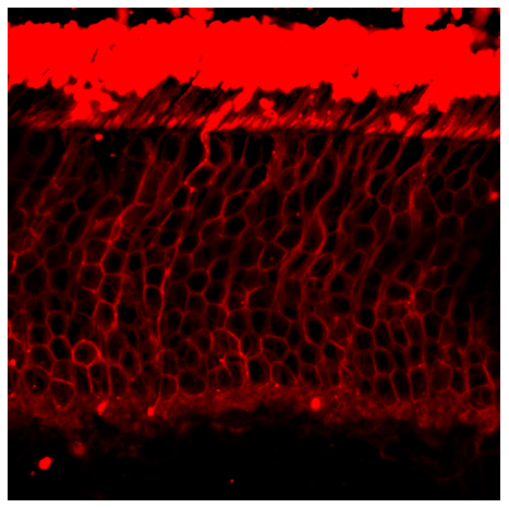
Images (1) Click the Picture to Zoom In
-
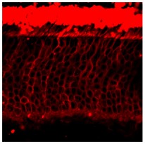
ARG52417 anti-Rhodopsin antibody [1D4] IHC image
Immunohistochemistry:Immunofluorescence: adult mouse retinal section stained with anti-rhodopsin antibody (ARG52417)
Clone References
Phagosome maturation during endosome interaction revealed by partial rhodopsin processing in retinal pigment epithelium.


Effective delivery of large genes to the retina by dual AAV vectors.
IHC-Fr / Mouse
Myosin7a deficiency results in reduced retinal activity which is improved by gene therapy.
EM / Mouse
CERKL knockdown causes retinal degeneration in zebrafish.
IHC-Fr / Zebrafish
Reproducible and sustained regulation of Gαs signalling using a metazoan opsin as an optogenetic tool.
ICC/IF / Human
Degeneration of cortical function in the Royal College of Surgeons rat.
IHC-FoFr / Rat
A neural-specific F-box protein Fbs1 functions as a chaperone suppressing glycoprotein aggregation.
IP / Human
Antigen-antibody interaction. Synthetic peptides define linear antigenic determinants recognized by monoclonal antibodies directed to the cytoplasmic carboxyl terminus of rhodopsin.
IP / Bovine


