ARG57488
anti-RhoA + RhoB + RhoC antibody
anti-RhoA + RhoB + RhoC antibody for Flow cytometry,ICC/IF,IHC-Formalin-fixed paraffin-embedded sections,Western blot and Human,Mouse,Rat
Overview
| Product Description | Rabbit Polyclonal antibody recognizes RhoA + RhoB + RhoC |
|---|---|
| Tested Reactivity | Hu, Ms, Rat |
| Tested Application | FACS, ICC/IF, IHC-P, WB |
| Specificity | This antibody detects endogenous levels of total RhoA + RhoB + RhoC. |
| Host | Rabbit |
| Clonality | Polyclonal |
| Isotype | IgG |
| Target Name | RhoA + RhoB + RhoC |
| Antigen Species | Human |
| Immunogen | Synthetic peptide derived from Human RhoA + RhoB + RhoC. |
| Conjugation | Un-conjugated |
| Alternate Names | Transforming protein RhoA; Rho cDNA clone 12; RHOH12; h12; ARHA; ARH12; RHO12 |
Application Instructions
| Application Suggestion |
|
||||||||||
|---|---|---|---|---|---|---|---|---|---|---|---|
| Application Note | * The dilutions indicate recommended starting dilutions and the optimal dilutions or concentrations should be determined by the scientist. |
Properties
| Form | Liquid |
|---|---|
| Purification | Purified by affinity chromatography. |
| Buffer | PBS (pH 7.4), 150mM NaCl, 0.02% Sodium azide and 50% Glycerol. |
| Preservative | 0.02% Sodium azide |
| Stabilizer | 50% Glycerol |
| Storage Instruction | For continuous use, store undiluted antibody at 2-8°C for up to a week. For long-term storage, aliquot and store at -20°C. Storage in frost free freezers is not recommended. Avoid repeated freeze/thaw cycles. Suggest spin the vial prior to opening. The antibody solution should be gently mixed before use. |
| Note | For laboratory research only, not for drug, diagnostic or other use. |
Bioinformation
| Database Links | |
|---|---|
| Gene Symbol | RHOA |
| Gene Full Name | ras homolog family member A |
| Background | This gene encodes a member of the Rho family of small GTPases, which cycle between inactive GDP-bound and active GTP-bound states and function as molecular switches in signal transduction cascades. Rho proteins promote reorganization of the actin cytoskeleton and regulate cell shape, attachment, and motility. Overexpression of this gene is associated with tumor cell proliferation and metastasis. Multiple alternatively spliced variants have been identified. [provided by RefSeq, Sep 2015] |
| Function | Regulates a signal transduction pathway linking plasma membrane receptors to the assembly of focal adhesions and actin stress fibers. Involved in a microtubule-dependent signal that is required for the myosin contractile ring formation during cell cycle cytokinesis. Plays an essential role in cleavage furrow formation. Required for the apical junction formation of keratinocyte cell-cell adhesion. Serves as a target for the yopT cysteine peptidase from Yersinia pestis, vector of the plague, and Yersinia pseudotuberculosis, which causes gastrointestinal disorders. Stimulates PKN2 kinase activity. May be an activator of PLCE1. Activated by ARHGEF2, which promotes the exchange of GDP for GTP. Essential for the SPATA13-mediated regulation of cell migration and adhesion assembly and disassembly. The MEMO1-RHOA-DIAPH1 signaling pathway plays an important role in ERBB2-dependent stabilization of microtubules at the cell cortex. It controls the localization of APC and CLASP2 to the cell membrane, via the regulation of GSK3B activity. In turn, membrane-bound APC allows the localization of the MACF1 to the cell membrane, which is required for microtubule capture and stabilization.Regulates a signal transduction pathway linking plasma membrane receptors to the assembly of focal adhesions and actin stress fibers. Involved in a microtubule-dependent signal that is required for the myosin contractile ring formation during cell cycle cytokinesis. Plays an essential role in cleavage furrow formation. Required for the apical junction formation of keratinocyte cell-cell adhesion. May be an activator of PLCE1. Activated by ARHGEF2, which promotes the exchange of GDP for GTP. Essential for the SPATA13-mediated regulation of cell migration and adhesion assembly and disassembly. The MEMO1-RHOA-DIAPH1 signaling pathway plays an important role in ERBB2-dependent stabilization of microtubules at the cell cortex. It controls the localization of APC and CLASP2 to the cell membrane, via the regulation of GSK3B activity. In turn, membrane-bound APC allows the localization of the MACF1 to the cell membrane, which is required for microtubule capture and stabilization (By similarity). Regulates KCNA2 potassium channel activity by reducing its location at the cell surface in response to CHRM1 activation; promotes KCNA2 endocytosis. [UniProt] |
| Calculated MW | 22 kDa |
| PTM | (Microbial infection) Substrate for botulinum ADP-ribosyltransferase. (Microbial infection) Cleaved by yopT protease when the cell is infected by some Yersinia pathogens. This removes the lipid attachment, and leads to its displacement from plasma membrane and to subsequent cytoskeleton cleavage. (Microbial infection) AMPylation at Tyr-34 and Thr-37 are mediated by bacterial enzymes in case of infection by H.somnus and V.parahaemolyticus, respectively. AMPylation occurs in the effector region and leads to inactivation of the GTPase activity by preventing the interaction with downstream effectors, thereby inhibiting actin assembly in infected cells. It is unclear whether some human enzyme mediates AMPylation; FICD has such ability in vitro but additional experiments remain to be done to confirm results in vivo. (Microbial infection) Glycosylated at Tyr-34 by Photorhabdus asymbiotica toxin PAU_02230. Mono-O-GlcNAcylation by PAU_02230 inhibits downstream signaling by an impaired interaction with diverse regulator and effector proteins of Rho and leads to actin disassembly. Phosphorylation by PRKG1 at Ser-188 inactivates RHOA signaling (PubMed:11162591). Phosphorylation by SLK at Ser-188 in response to AGTR2 activation (By similarity). Ubiquitinated by the BCR(BACURD1) and BCR(BACURD2) E3 ubiquitin ligase complexes, leading to its degradation by the proteasome, thereby regulating the actin cytoskeleton and cell migration. |
Images (3) Click the Picture to Zoom In
-

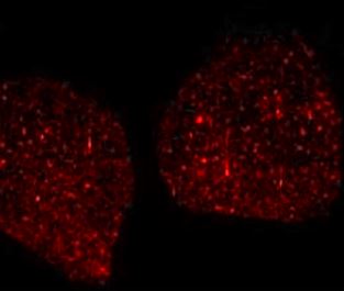
ARG57488 anti-RhoA + RhoB + RhoC antibody ICC/IF image
Immunofluorescence: HeLa cells stained with ARG57488 anti-RhoA + RhoB + RhoC antibody.
-
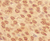
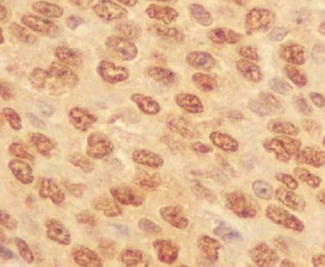
ARG57488 anti-RhoA + RhoB + RhoC antibody IHC-P image
Immunohistochemistry: Paraffin-embedded Human lung carcinoma stained with ARG57488 anti-RhoA + RhoB + RhoC antibody.
-
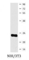
ARG57488 anti-RhoA + RhoB + RhoC antibody WB image
Western blot: NIH/3T3 cell lysate stained with ARG57488 anti-RhoA + RhoB + RhoC antibody.