ARG43056
anti-Ret antibody
anti-Ret antibody for IHC-Formalin-fixed paraffin-embedded sections and Human,Mouse,Rat
Overview
| Product Description | Rabbit Polyclonal antibody recognizes Ret |
|---|---|
| Tested Reactivity | Hu, Ms, Rat |
| Tested Application | IHC-P |
| Host | Rabbit |
| Clonality | Polyclonal |
| Isotype | IgG |
| Target Name | Ret |
| Antigen Species | Human |
| Immunogen | Recombinant protein corresponding to L29-V304 of Human Ret. |
| Conjugation | Un-conjugated |
| Alternate Names | RET51; CDHF12; HSCR1; Proto-oncogene c-Ret; PTC; Proto-oncogene tyrosine-protein kinase receptor Ret; RET-ELE1; CDHR16; MEN2B; MEN2A; MTC1; EC 2.7.10.1; Cadherin family member 12 |
Application Instructions
| Application Suggestion |
|
||||
|---|---|---|---|---|---|
| Application Note | IHC-P: Antigen Retrieval: Heat mediation was performed in Citrate buffer (pH 6.0) for 20 min. * The dilutions indicate recommended starting dilutions and the optimal dilutions or concentrations should be determined by the scientist. |
Properties
| Form | Liquid |
|---|---|
| Purification | Affinity purification with immunogen. |
| Buffer | 0.2% Na2HPO4, 0.9% NaCl, 0.05% Sodium azide and 4% Trehalose. |
| Preservative | 0.05% Sodium azide |
| Stabilizer | 4% Trehalose |
| Concentration | 0.5 mg/ml |
| Storage Instruction | For continuous use, store undiluted antibody at 2-8°C for up to a week. For long-term storage, aliquot and store at -20°C or below. Storage in frost free freezers is not recommended. Avoid repeated freeze/thaw cycles. Suggest spin the vial prior to opening. The antibody solution should be gently mixed before use. |
| Note | For laboratory research only, not for drug, diagnostic or other use. |
Bioinformation
| Database Links | |
|---|---|
| Gene Symbol | RET |
| Gene Full Name | ret proto-oncogene |
| Background | This gene encodes a transmembrane receptor and member of the tyrosine protein kinase family of proteins. Binding of ligands such as GDNF (glial cell-line derived neurotrophic factor) and other related proteins to the encoded receptor stimulates receptor dimerization and activation of downstream signaling pathways that play a role in cell differentiation, growth, migration and survival. The encoded receptor is important in development of the nervous system, and the development of organs and tissues derived from the neural crest. This proto-oncogene can undergo oncogenic activation through both cytogenetic rearrangement and activating point mutations. Mutations in this gene are associated with Hirschsprung disease and central hypoventilation syndrome and have been identified in patients with renal agenesis. [provided by RefSeq, Sep 2017] |
| Function | Receptor tyrosine-protein kinase involved in numerous cellular mechanisms including cell proliferation, neuronal navigation, cell migration, and cell differentiation upon binding with glial cell derived neurotrophic factor family ligands. Phosphorylates PTK2/FAK1. Regulates both cell death/survival balance and positional information. Required for the molecular mechanisms orchestration during intestine organogenesis; involved in the development of enteric nervous system and renal organogenesis during embryonic life, and promotes the formation of Peyer's patch-like structures, a major component of the gut-associated lymphoid tissue. Modulates cell adhesion via its cleavage by caspase in sympathetic neurons and mediates cell migration in an integrin (e.g. ITGB1 and ITGB3)-dependent manner. Involved in the development of the neural crest. Active in the absence of ligand, triggering apoptosis through a mechanism that requires receptor intracellular caspase cleavage. Acts as a dependence receptor; in the presence of the ligand GDNF in somatotrophs (within pituitary), promotes survival and down regulates growth hormone (GH) production, but triggers apoptosis in absence of GDNF. Regulates nociceptor survival and size. Triggers the differentiation of rapidly adapting (RA) mechanoreceptors. Mediator of several diseases such as neuroendocrine cancers; these diseases are characterized by aberrant integrins-regulated cell migration. Mediates, through interaction with GDF15-receptor GFRAL, GDF15-induced cell-signaling in the brainstem which induces inhibition of food-intake. Activates MAPK- and AKT-signaling pathways (PubMed:28846097, PubMed:28953886, PubMed:28846099). Isoform 1 in complex with GFRAL induces higher activation of MAPK-signaling pathway than isoform 2 in complex with GFRAL (PubMed:28846099). [UniProt] |
| Cellular Localization | Cell membrane; Single-pass type I membrane protein. Endosome membrane; Single-pass type I membrane protein. [UniProt] |
| Calculated MW | 124 kDa |
| PTM | Autophosphorylated on C-terminal tyrosine residues upon ligand stimulation. Dephosphorylated by PTPRJ on Tyr-905, Tyr-1015 and Tyr-1062. Proteolytically cleaved by caspase-3. The soluble RET kinase fragment is able to induce cell death. The extracellular cell-membrane anchored RET cadherin fragment accelerates cell adhesion in sympathetic neurons. [UniProt] |
Images (6) Click the Picture to Zoom In
-

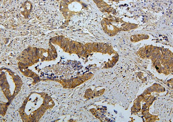
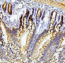
ARG43056 anti-Ret antibody IHC-P image
Immunohistochemistry: Paraffin-embedded Human colon cancer tissue. Antigen Retrieval: Heat mediation was performed in Citrate buffer (pH 6.0) for 20 min. The tissue section was blocked with 10% goat serum. The tissue section was then stained with ARG43056 anti-Ret antibody at 1 µg/ml dilution, overnight at 4°C.
-
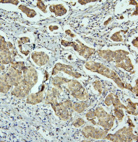
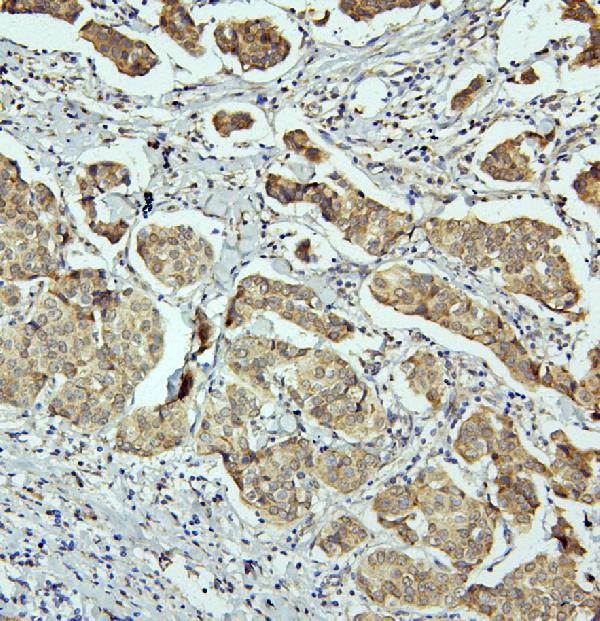
ARG43056 anti-Ret antibody IHC-P image
Immunohistochemistry: Paraffin-embedded Human mammary cancer tissue. Antigen Retrieval: Heat mediation was performed in Citrate buffer (pH 6.0) for 20 min. The tissue section was blocked with 10% goat serum. The tissue section was then stained with ARG43056 anti-Ret antibody at 1 µg/ml dilution, overnight at 4°C.
-
ARG43056 anti-Ret antibody IHC-P image
Immunohistochemistry: Paraffin-embedded Mouse intestine tissue. Antigen Retrieval: Heat mediation was performed in Citrate buffer (pH 6.0) for 20 min. The tissue section was blocked with 10% goat serum. The tissue section was then stained with ARG43056 anti-Ret antibody at 1 µg/ml dilution, overnight at 4°C.
-
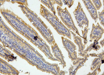
ARG43056 anti-Ret antibody IHC-P image
Immunohistochemistry: Paraffin-embedded Rat intestine tissue. Antigen Retrieval: Heat mediation was performed in Citrate buffer (pH 6.0) for 20 min. The tissue section was blocked with 10% goat serum. The tissue section was then stained with ARG43056 anti-Ret antibody at 1 µg/ml dilution, overnight at 4°C.
-

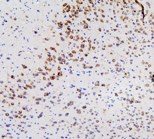
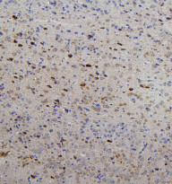
ARG43056 anti-Ret antibody IHC-P image
Immunohistochemistry: Paraffin-embedded Mouse brain tissue. Antigen Retrieval: Heat mediation was performed in Citrate buffer (pH 6.0) for 20 min. The tissue section was blocked with 10% goat serum. The tissue section was then stained with ARG43056 anti-Ret antibody at 1 µg/ml dilution, overnight at 4°C.
-
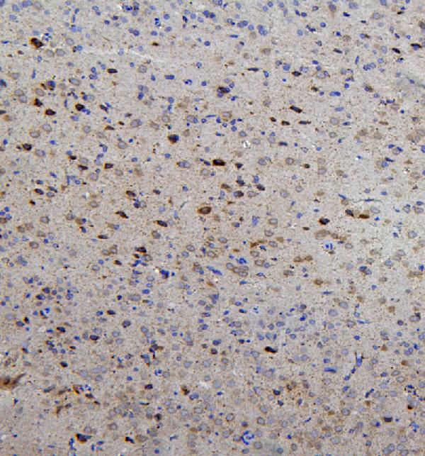
ARG43056 anti-Ret antibody IHC-P image
Immunohistochemistry: Paraffin-embedded Rat brain tissue. Antigen Retrieval: Heat mediation was performed in Citrate buffer (pH 6.0) for 20 min. The tissue section was blocked with 10% goat serum. The tissue section was then stained with ARG43056 anti-Ret antibody at 1 µg/ml dilution, overnight at 4°C.