ARG40999
anti-Ran antibody [5D5]
anti-Ran antibody [5D5] for Flow cytometry,ICC/IF,Western blot and Human,Mouse,Rat
Overview
| Product Description | Mouse Monoclonal antibody [5D5] recognizes Ran |
|---|---|
| Tested Reactivity | Hu, Ms, Rat |
| Tested Application | FACS, ICC/IF, WB |
| Host | Mouse |
| Clonality | Monoclonal |
| Clone | 5D5 |
| Isotype | IgG2b |
| Target Name | Ran |
| Antigen Species | Human |
| Immunogen | Recombinant protein corresponding to A2-L216 of Human Ran. |
| Conjugation | Un-conjugated |
| Alternate Names | GTP-binding nuclear protein Ran; Androgen receptor-associated protein 24; TC4; ARA24; Ras-like protein TC4; Gsp1; Ras-related nuclear protein; GTPase Ran |
Application Instructions
| Application Suggestion |
|
||||||||
|---|---|---|---|---|---|---|---|---|---|
| Application Note | * The dilutions indicate recommended starting dilutions and the optimal dilutions or concentrations should be determined by the scientist. | ||||||||
| Observed Size | 24 kDa |
Properties
| Form | Liquid |
|---|---|
| Purification | Affinity purification with immunogen. |
| Buffer | 0.2% Na2HPO4, 0.9% NaCl, 0.05% Sodium azide and 4% Trehalose. |
| Preservative | 0.05% Sodium azide |
| Stabilizer | 4% Trehalose |
| Concentration | 0.5 mg/ml |
| Storage Instruction | For continuous use, store undiluted antibody at 2-8°C for up to a week. For long-term storage, aliquot and store at -20°C or below. Storage in frost free freezers is not recommended. Avoid repeated freeze/thaw cycles. Suggest spin the vial prior to opening. The antibody solution should be gently mixed before use. |
| Note | For laboratory research only, not for drug, diagnostic or other use. |
Bioinformation
| Database Links | |
|---|---|
| Gene Symbol | RAN |
| Gene Full Name | RAN, member RAS oncogene family |
| Background | RAN (ras-related nuclear protein) is a small GTP binding protein belonging to the RAS superfamily that is essential for the translocation of RNA and proteins through the nuclear pore complex. The RAN protein is also involved in control of DNA synthesis and cell cycle progression. Nuclear localization of RAN requires the presence of regulator of chromosome condensation 1 (RCC1). Mutations in RAN disrupt DNA synthesis. Because of its many functions, it is likely that RAN interacts with several other proteins. RAN regulates formation and organization of the microtubule network independently of its role in the nucleus-cytosol exchange of macromolecules. RAN could be a key signaling molecule regulating microtubule polymerization during mitosis. RCC1 generates a high local concentration of RAN-GTP around chromatin which, in turn, induces the local nucleation of microtubules. RAN is an androgen receptor (AR) coactivator that binds differentially with different lengths of polyglutamine within the androgen receptor. Polyglutamine repeat expansion in the AR is linked to Kennedy's disease (X-linked spinal and bulbar muscular atrophy). RAN coactivation of the AR diminishes with polyglutamine expansion within the AR, and this weak coactivation may lead to partial androgen insensitivity during the development of Kennedy's disease. [provided by RefSeq, Jul 2008] |
| Function | GTP-binding protein involved in nucleocytoplasmic transport. Required for the import of protein into the nucleus and also for RNA export. Involved in chromatin condensation and control of cell cycle (By similarity). The complex with BIRC5/ survivin plays a role in mitotic spindle formation by serving as a physical scaffold to help deliver the RAN effector molecule TPX2 to microtubules. Acts as a negative regulator of the kinase activity of VRK1 and VRK2. Enhances AR-mediated transactivation. Transactivation decreases as the poly-Gln length within AR increases. [UniProt] |
| Cellular Localization | Nucleus. Nucleus envelope. Cytoplasm, cytosol. Cytoplasm. Melanosome. Note=Predominantly nuclear during interphase (PubMed:8421051, PubMed:12194828, PubMed:10679025). Becomes dispersed throughout the cytoplasm during mitosis (PubMed:8421051, PubMed:12194828). Identified by mass spectrometry in melanosome fractions from stage I to stage IV (PubMed:17081065). [UniProt] |
| Calculated MW | 24 kDa |
Images (4) Click the Picture to Zoom In
-
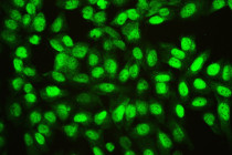
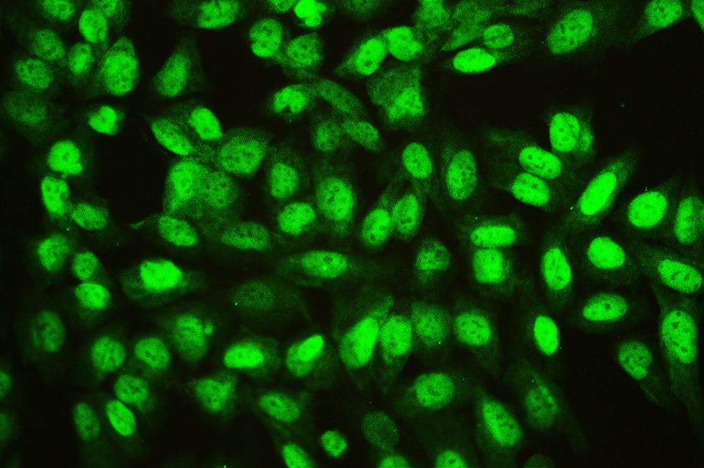
ARG40999 anti-Ran antibody [5D5] ICC/IF image
Immunofluorescence: U2OS cells stained with ARG40999 anti-Ran antibody [5D5] at 2 µg/ml, overnight at 4°C.
-
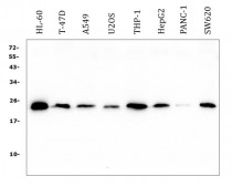
ARG40999 anti-Ran antibody [5D5] WB image
Western blot: 50 µg of samples under reducing conditions. HL-60, T-47D, A549, U2OS, THP-1, HepG2, PANC-1 and SW620 whole cell lysates stained with ARG40999 anti-Ran antibody [5D5] at 0.5 µg/ml, overnight at 4°C.
-

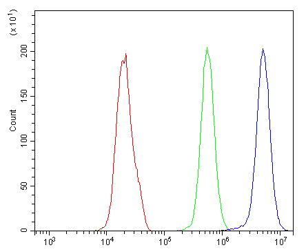
ARG40999 anti-Ran antibody [5D5] FACS image
Flow Cytometry: PC-3 cells were blocked with 10% normal goat serum and then stained with ARG40999 anti-Ran antibody [5D5] (blue) at 1 µg/10^6 cells for 30 min at 20°C, followed by incubation with DyLight®488 labelled secondary antibody. Isotype control antibody (green) was Mouse IgG (1 µg/10^6 cells) used under the same conditions. Unlabelled sample (red) was also used as a control.
-

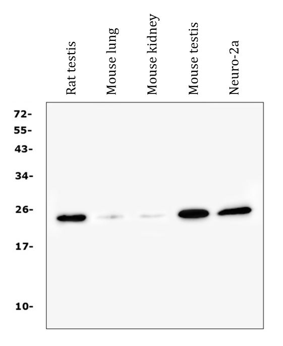
ARG40999 anti-Ran antibody [5D5] WB image
Western blot: 50 µg of samples under reducing conditions. Rat testis, Mouse lung, Mouse kidney, Mouse testis and Mouse Neuro-2a whole cell lysates stained with ARG40999 anti-Ran antibody [5D5] at 0.5 µg/ml, overnight at 4°C.