ARG42689
anti-RUNX3 antibody
anti-RUNX3 antibody for Flow cytometry,ICC/IF,IHC-Formalin-fixed paraffin-embedded sections,Western blot and Human,Mouse,Rat
Overview
| Product Description | Rabbit Polyclonal antibody recognizes RUNX3 |
|---|---|
| Tested Reactivity | Hu, Ms, Rat |
| Tested Application | FACS, ICC/IF, IHC-P, WB |
| Host | Rabbit |
| Clonality | Polyclonal |
| Isotype | IgG |
| Target Name | RUNX3 |
| Antigen Species | Human |
| Immunogen | Recombinant protein corresponding to M128-Y270 of Human RUNX3. |
| Conjugation | Un-conjugated |
| Alternate Names | Runt-related transcription factor 3; AML2; Polyomavirus enhancer-binding protein 2 alpha C subunit; CBFA3; SL3/AKV core-binding factor alpha C subunit; Oncogene AML-2; SL3-3 enhancer factor 1 alpha C subunit; PEBP2aC; Core-binding factor subunit alpha-3; CBF-alpha-3; PEA2-alpha C; PEBP2-alpha C; Acute myeloid leukemia 2 protein |
Application Instructions
| Application Suggestion |
|
||||||||||
|---|---|---|---|---|---|---|---|---|---|---|---|
| Application Note | * The dilutions indicate recommended starting dilutions and the optimal dilutions or concentrations should be determined by the scientist. | ||||||||||
| Positive Control | A431 and U2OS | ||||||||||
| Observed Size | ~ 44 kDa |
Properties
| Form | Liquid |
|---|---|
| Purification | Affinity purification with immunogen. |
| Buffer | 0.2% Na2HPO4, 0.9% NaCl, 0.05% Sodium azide and 5% BSA. |
| Preservative | 0.05% Sodium azide |
| Stabilizer | 5% BSA |
| Concentration | 0.5 mg/ml |
| Storage Instruction | For continuous use, store undiluted antibody at 2-8°C for up to a week. For long-term storage, aliquot and store at -20°C or below. Storage in frost free freezers is not recommended. Avoid repeated freeze/thaw cycles. Suggest spin the vial prior to opening. The antibody solution should be gently mixed before use. |
| Note | For laboratory research only, not for drug, diagnostic or other use. |
Bioinformation
| Database Links |
Swiss-port # Q13761 Human Runt-related transcription factor 3 |
|---|---|
| Gene Symbol | RUNX3 |
| Gene Full Name | runt-related transcription factor 3 |
| Background | This gene encodes a member of the runt domain-containing family of transcription factors. A heterodimer of this protein and a beta subunit forms a complex that binds to the core DNA sequence 5'-PYGPYGGT-3' found in a number of enhancers and promoters, and can either activate or suppress transcription. It also interacts with other transcription factors. It functions as a tumor suppressor, and the gene is frequently deleted or transcriptionally silenced in cancer. Alternative splicing results in multiple transcript variants. [provided by RefSeq, Mar 2016] |
| Function | Forms the heterodimeric complex core-binding factor (CBF) with CBFB. RUNX members modulate the transcription of their target genes through recognizing the core consensus binding sequence 5'-TGTGGT-3', or very rarely, 5'-TGCGGT-3', within their regulatory regions via their runt domain, while CBFB is a non-DNA-binding regulatory subunit that allosterically enhances the sequence-specific DNA-binding capacity of RUNX. The heterodimers bind to the core site of a number of enhancers and promoters, including murine leukemia virus, polyomavirus enhancer, T-cell receptor enhancers, LCK, IL3 and GM-CSF promoters (By similarity). May be involved in the control of cellular proliferation and/or differentiation. In association with ZFHX3, upregulates CDKN1A promoter activity following TGF-beta stimulation (PubMed:20599712). CBF complexes repress ZBTB7B transcription factor during cytotoxic (CD8+) T cell development. They bind to RUNX-binding sequence within the ZBTB7B locus acting as transcriptional silencer and allowing for cytotoxic T cell differentiation. CBF complexes binding to the transcriptional silencer is essential for recruitment of nuclear protein complexes that catalyze epigenetic modifications to establish epigenetic ZBTB7B silencing (By similarity). [UniProt] |
| Cellular Localization | Nucleus. Cytoplasm. Note=The tyrosine phosphorylated form localizes to the cytoplasm. Translocates from the cytoplasm to the nucleus following TGF-beta stimulation. [UniProt] |
| Calculated MW | 44 kDa |
| PTM | Phosphorylated on tyrosine residues by SRC. Phosphorylated by LCK and FYN. [UniProt] |
Images (7) Click the Picture to Zoom In
-
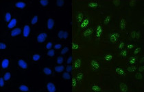
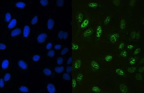
ARG42689 anti-RUNX3 antibody ICC/IF image
Immunofluorescence: U2OS cells were blocked with 10% goat serum and then stained with ARG42689 anti-RUNX3 antibody (green) at 2 µg/ml dilution, overnight at 4°C. DAPI (blue) for nuclear staining.
-
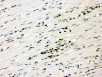
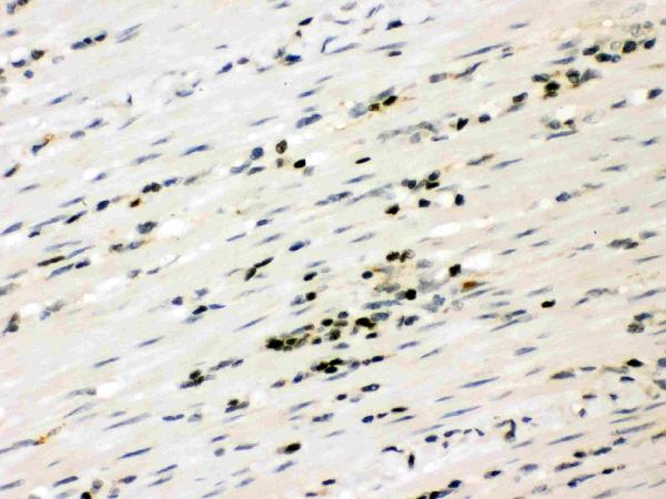
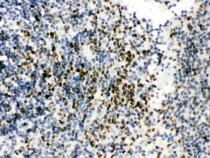
ARG42689 anti-RUNX3 antibody IHC-P image
Immunohistochemistry: Paraffin-embedded Human intestinal cancer tissue stained with ARG42689 anti-RUNX3 antibody.
-
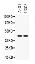
ARG42689 anti-RUNX3 antibody WB image
Western blot: 40 µg of A431 and U2OS whole cell lysates stained with ARG42689 anti-RUNX3 antibody at 0.5 µg/ml dilution.
-
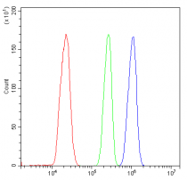
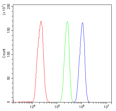
ARG42689 anti-RUNX3 antibody FACS image
Flow Cytometry: THP-1 cells were blocked with 10% normal goat serum and then stained with ARG42689 anti-RUNX3 antibody (blue) at 1 µg/10^6 cells for 30 min at 20°C, followed by incubation with DyLight®488 labelled secondary antibody. Isotype control antibody (green) was rabbit IgG (1 µg/10^6 cells) used under the same conditions. Unlabelled sample (red) was also used as a control.
-
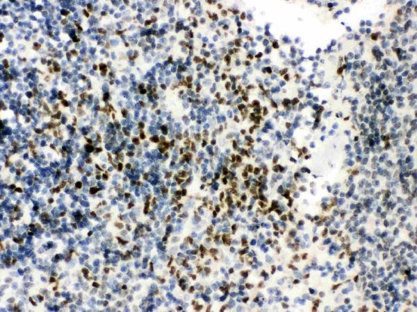
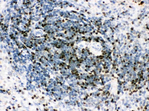
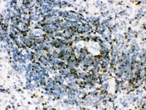
ARG42689 anti-RUNX3 antibody IHC-P image
Immunohistochemistry: Paraffin-embedded Mouse spleen tissue stained with ARG42689 anti-RUNX3 antibody.
-
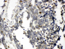
ARG42689 anti-RUNX3 antibody IHC-P image
Immunohistochemistry: Paraffin-embedded Rat spleen tissue stained with ARG42689 anti-RUNX3 antibody.
-
ARG42689 anti-RUNX3 antibody IHC-P image
Immunohistochemistry: Paraffin-embedded Human lung cancer tissue stained with ARG42689 anti-RUNX3 antibody.