ARG42970
anti-RP2 / XRP2 antibody [3D7]
anti-RP2 / XRP2 antibody [3D7] for Flow cytometry,ICC/IF,Western blot and Human
Overview
| Product Description | Mouse Monoclonal antibody [3D7] recognizes RP2 / XRP2 |
|---|---|
| Tested Reactivity | Hu |
| Tested Application | FACS, ICC/IF, WB |
| Host | Mouse |
| Clonality | Monoclonal |
| Clone | 3D7 |
| Isotype | IgG2b |
| Target Name | RP2 / XRP2 |
| Antigen Species | Human |
| Immunogen | Recombinant protein corresponding to D244-M348 of Human RP2 / XRP2. |
| Conjugation | Un-conjugated |
| Alternate Names | TBCCD2; Protein XRP2; XRP2; DELXp11.3; NM23-H10; NME10 |
Application Instructions
| Application Suggestion |
|
||||||||
|---|---|---|---|---|---|---|---|---|---|
| Application Note | * The dilutions indicate recommended starting dilutions and the optimal dilutions or concentrations should be determined by the scientist. | ||||||||
| Observed Size | ~ 40 kDa |
Properties
| Form | Liquid |
|---|---|
| Purification | Affinity purification with immunogen. |
| Buffer | 0.2% Na2HPO4, 0.9% NaCl, 0.05% Sodium azide and 4% Trehalose. |
| Preservative | 0.05% Sodium azide |
| Stabilizer | 4% Trehalose |
| Concentration | 0.5 mg/ml |
| Storage Instruction | For continuous use, store undiluted antibody at 2-8°C for up to a week. For long-term storage, aliquot and store at -20°C or below. Storage in frost free freezers is not recommended. Avoid repeated freeze/thaw cycles. Suggest spin the vial prior to opening. The antibody solution should be gently mixed before use. |
| Note | For laboratory research only, not for drug, diagnostic or other use. |
Bioinformation
| Database Links | |
|---|---|
| Gene Symbol | RP2 |
| Gene Full Name | retinitis pigmentosa 2 (X-linked recessive) |
| Background | The RP2 locus has been implicated as one cause of X-linked retinitis pigmentosa. The predicted gene product shows homology with human cofactor C, a protein involved in the ultimate step of beta-tubulin folding. Progressive retinal degeneration may therefore be due to the accumulation of incorrectly-folded photoreceptor or neuron-specific tubulin isoforms followed by progressive cell death [provided by RefSeq, Jul 2008] |
| Function | Acts as a GTPase-activating protein (GAP) involved in trafficking between the Golgi and the ciliary membrane. Involved in localization of proteins, such as NPHP3, to the cilium membrane by inducing hydrolysis of GTP ARL3, leading to the release of UNC119 (or UNC119B). Acts as a GTPase-activating protein (GAP) for tubulin in concert with tubulin-specific chaperone C, but does not enhance tubulin heterodimerization. Acts as guanine nucleotide dissociation inhibitor towards ADP-ribosylation factor-like proteins. [UniProt] |
| Cellular Localization | Cell membrane; Lipid-anchor; Cytoplasmic side. Cell projection, cilium. Note=Detected predominantly at the plasma membrane of rod and cone photoreceptors. Not detected in the nucleus. [UniProt] |
| Calculated MW | 40 kDa |
| PTM | Myristoylated on Gly-2; which may be required for membrane targeting. Palmitoylated on Cys-3; which may be required for plasma membrane targeting (Probable). Mutation of Cys-3 targets the protein to internal membranes. [UniProt] |
Images (3) Click the Picture to Zoom In
-
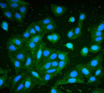
ARG42970 anti-RP2 / XRP2 antibody [3D7] ICC/IF image
Immunofluorescence: U2OS stained with ARG42970 anti-RP2 / XRP2 antibody [3D7] at 2 μg/mL dilution.
-

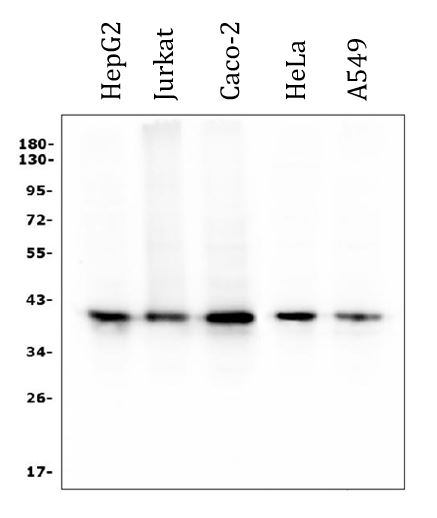
ARG42970 anti-RP2 / XRP2 antibody [3D7] WB image
Western blot: 50 µg of sample under reducing conditions. HepG2, Jurkat, Caco-2, HeLa and A549 whole cell lysates stained with ARG42970 anti-RP2 / XRP2 antibody [3D7] at 0.5 µg/ml dilution, overnight at 4°C.
-
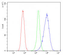
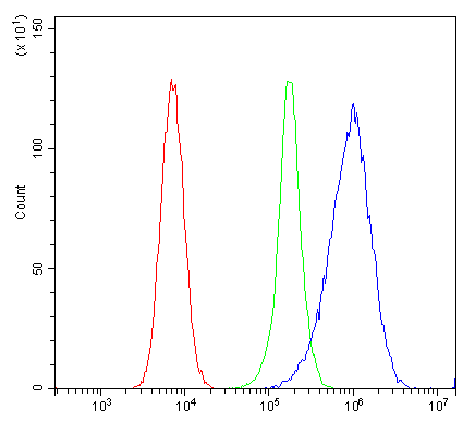
ARG42970 anti-RP2 / XRP2 antibody [3D7] FACS image
Flow Cytometry: 293T cells were blocked with 10% normal goat serum and then stained with ARG42970 anti-RP2 / XRP2 antibody [3D7] (blue) at 1 µg/10^6 cells for 30 min at 20°C, followed by incubation with DyLight®488 labelled secondary antibody. Isotype control antibody (green) was Mouse IgG (1 µg/10^6 cells) used under the same conditions. Unlabelled sample (red) was also used as a control.