ARG43232
anti-RICTOR antibody
anti-RICTOR antibody for Flow cytometry,ICC/IF,IHC-Formalin-fixed paraffin-embedded sections,Western blot and Human
Overview
| Product Description | Rabbit Polyclonal antibody recognizes RICTOR |
|---|---|
| Tested Reactivity | Hu |
| Tested Application | FACS, ICC/IF, IHC-P, WB |
| Host | Rabbit |
| Clonality | Polyclonal |
| Isotype | IgG |
| Target Name | RICTOR |
| Antigen Species | Human |
| Immunogen | Recombinant protein corresponding to E23-H264 of Human RICTOR. |
| Conjugation | Un-conjugated |
| Alternate Names | AVO3 homolog; PIA; Rapamycin-insensitive companion of mTOR; hAVO3; AVO3 |
Application Instructions
| Application Suggestion |
|
||||||||||
|---|---|---|---|---|---|---|---|---|---|---|---|
| Application Note | IHC-P: Antigen Retrieval: Heat mediation was performed in EDTA buffer (pH 8.0). * The dilutions indicate recommended starting dilutions and the optimal dilutions or concentrations should be determined by the scientist. |
||||||||||
| Observed Size | ~ 210 kDa |
Properties
| Form | Liquid |
|---|---|
| Purification | Affinity purification with immunogen. |
| Buffer | 0.2% Na2HPO4, 0.9% NaCl, 0.05% Sodium azide and 4% Trehalose. |
| Preservative | 0.05% Sodium azide |
| Stabilizer | 4% Trehalose |
| Concentration | 0.5 mg/ml |
| Storage Instruction | For continuous use, store undiluted antibody at 2-8°C for up to a week. For long-term storage, aliquot and store at -20°C or below. Storage in frost free freezers is not recommended. Avoid repeated freeze/thaw cycles. Suggest spin the vial prior to opening. The antibody solution should be gently mixed before use. |
| Note | For laboratory research only, not for drug, diagnostic or other use. |
Bioinformation
| Database Links |
Swiss-port # Q6R327 Human Rapamycin-insensitive companion of mTOR |
|---|---|
| Gene Symbol | RICTOR |
| Gene Full Name | RPTOR independent companion of MTOR, complex 2 |
| Background | RICTOR and MTOR (FRAP1; MIM 601231) are components of a protein complex that integrates nutrient- and growth factor-derived signals to regulate cell growth (Sarbassov et al., 2004 [PubMed 15268862]).[supplied by OMIM, Mar 2008] |
| Function | Subunit of mTORC2, which regulates cell growth and survival in response to hormonal signals. mTORC2 is activated by growth factors, but, in contrast to mTORC1, seems to be nutrient-insensitive. mTORC2 seems to function upstream of Rho GTPases to regulate the actin cytoskeleton, probably by activating one or more Rho-type guanine nucleotide exchange factors. mTORC2 promotes the serum-induced formation of stress-fibers or F-actin. mTORC2 plays a critical role in AKT1 'Ser-473' phosphorylation, which may facilitate the phosphorylation of the activation loop of AKT1 on 'Thr-308' by PDK1 which is a prerequisite for full activation. mTORC2 regulates the phosphorylation of SGK1 at 'Ser-422'. mTORC2 also modulates the phosphorylation of PRKCA on 'Ser-657'. Plays an essential role in embryonic growth and development. [UniProt] |
| Calculated MW | 192 kDa |
| PTM | Phosphorylated by MTOR; when part of mTORC2. Phosphorylated at Thr-1135 by RPS6KB1; phosphorylation of RICTOR inhibits mTORC2 and AKT1 signaling. [UniProt] |
Images (5) Click the Picture to Zoom In
-
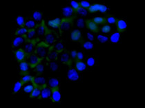
ARG43232 anti-RICTOR antibody ICC/IF image
Immunofluorescence: A431 cells stained with ARG43232 anti-RICTOR antibody (green) at 2 µg/ml dilution, overnight at 4°C. DAPI (blue) for nuclear staining.
-
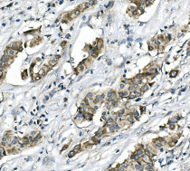
ARG43232 anti-RICTOR antibody IHC-P image
Immunohistochemistry: Paraffin-embedded Human mammary cancer tissue. Antigen Retrieval: Heat mediation was performed in EDTA buffer (pH 8.0). The tissue section was blocked with 10% goat serum. The tissue section was then stained with ARG43232 anti-RICTOR antibody at 1 µg/ml dilution, overnight at 4°C.
-
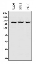
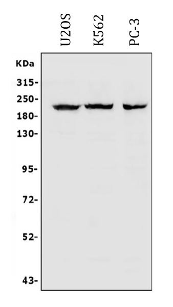
ARG43232 anti-RICTOR antibody WB image
Western blot: 30 µg of sample under reducing conditions. U2OS, K562 and PC-3 whole cell lysates stained with ARG43232 anti-RICTOR antibody at 0.5 µg/ml dilution, overnight at 4°C.
-
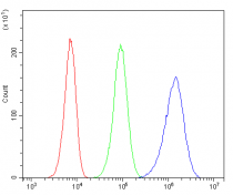
ARG43232 anti-RICTOR antibody FACS image
Flow Cytometry: 293T cells were blocked with 10% normal goat serum and then stained with ARG43232 anti-RICTOR antibody (blue) at 1 µg/10^6 cells for 30 min at 20°C, followed by incubation with DyLight®488 labelled secondary antibody. Isotype control antibody (green) was rabbit IgG (1 µg/10^6 cells) used under the same conditions. Unlabelled sample (red) was also used as a control.
-
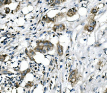
ARG43232 anti-RICTOR antibody IHC-P image
Immunohistochemistry: Paraffin-embedded Human mammary cancer tissue. Antigen Retrieval: Heat mediation was performed in EDTA buffer (pH 8.0). The tissue section was blocked with 10% goat serum. The tissue section was then stained with ARG43232 anti-RICTOR antibody at 1 µg/ml dilution, overnight at 4°C.